 Explore
 Validate
 Learn
Learn Western blot
 ELISA
ELISA Immunohistochemistry
Antibody data
- Antibody Data
- Antigen structure
- References [4]
- Comments [0]
- Validations
- Immunohistochemistry [1]
- Other assay [4]
Submit
Validation data
Reference
Comment
Report error
- Product number
- PA5-72765 - Provider product page

- Provider
- Invitrogen Antibodies
- Product name
- SIGLEC15 Polyclonal Antibody
- Antibody type
- Polyclonal
- Antigen
- Synthetic peptide
- Reactivity
- Human, Mouse, Rat
- Host
- Rabbit
- Isotype
- IgG
- Vial size
- 100 μg
- Concentration
- 1 mg/mL
- Storage
- Store at 4°C short term. For long term storage, store at -20°C, avoiding freeze/thaw cycles.
Submitted references Gene Expression Profiles Analyzed Using Integrating RNA Sequencing, and Microarray Reveals Increased Inflammatory Response, Proliferation, and Osteoclastogenesis in Pigmented Villonodular Synovitis.
Sialic acid-binding immunoglobulin-like lectin-15 expression on peritumoral macrophages is a favorable prognostic factor for primary central nervous system lymphoma patients.
The significance of Siglec-15 expression in resectable non-small cell lung cancer.
Expression signature, prognosis value, and immune characteristics of Siglec-15 identified by pan-cancer analysis.
Zhao Y, Lv J, Zhang H, Xie J, Dai H, Zhang X
Frontiers in immunology 2021;12:665442
Frontiers in immunology 2021;12:665442
Sialic acid-binding immunoglobulin-like lectin-15 expression on peritumoral macrophages is a favorable prognostic factor for primary central nervous system lymphoma patients.
Fudaba H, Momii Y, Hirakawa T, Onishi K, Asou D, Matsushita W, Kawasaki Y, Sugita K, Fujiki M
Scientific reports 2021 Jan 13;11(1):1206
Scientific reports 2021 Jan 13;11(1):1206
The significance of Siglec-15 expression in resectable non-small cell lung cancer.
Hao JQ, Nong JY, Zhao D, Li HY, Su D, Zhou LJ, Dong YJ, Zhang C, Che NY, Zhang SC, Lin JZ, Yang JB, Zhang HT, Wang JH
Neoplasma 2020 Nov;67(6):1214-1222
Neoplasma 2020 Nov;67(6):1214-1222
Expression signature, prognosis value, and immune characteristics of Siglec-15 identified by pan-cancer analysis.
Li B, Zhang B, Wang X, Zeng Z, Huang Z, Zhang L, Wei F, Ren X, Yang L
Oncoimmunology 2020 Aug 28;9(1):1807291
Oncoimmunology 2020 Aug 28;9(1):1807291
No comments: Submit comment
Supportive validation
- Submitted by
- Invitrogen Antibodies (provider)
- Main image
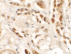
- Experimental details
- Immunohistochemistry of SIGLEC15 in human kidney tissue with SIGLEC15 Polyclonal Antibody (Product # PA5-72765) at 2.5 µg/mL.
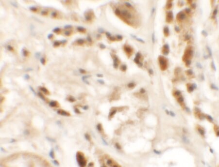
Supportive validation
- Submitted by
- Invitrogen Antibodies (provider)
- Main image
- Experimental details
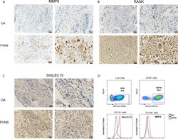
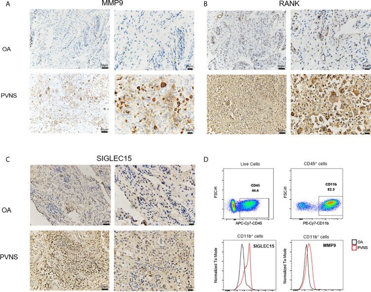
- Figure 7 Increased MMP9, SIGLEC 15, and RANK in PVNS patients MMP9 (A) , SIGLEC 15 (B) , and RANK (C) expression were detected by immunohistochemical test in synovium of PVNS and OA. Label is 50mum or 20mum. (D) SIGLEC 15 and MMP9 expression in surface of myeloid cell (CD45 + CD11b + ) were detected by flow cytometry.
- Submitted by
- Invitrogen Antibodies (provider)
- Main image
- Experimental details
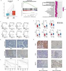
- Figure 6. Preliminary experimental verification of Siglec-15 signature in LUAD. (a) Siglec-15 expression on 20 paired LUAD tissues and adjacent normal tissues by RT-qPCR. (b) ""Chemokine signaling pathway"" associated with Siglec-15 in LUAD. ""Cytokine production"" and ""regulation of cytokine production"" included in the TOP20 GO terms in LUAD (NES >= 1.5, FDR

- Submitted by
- Invitrogen Antibodies (provider)
- Main image

- Experimental details
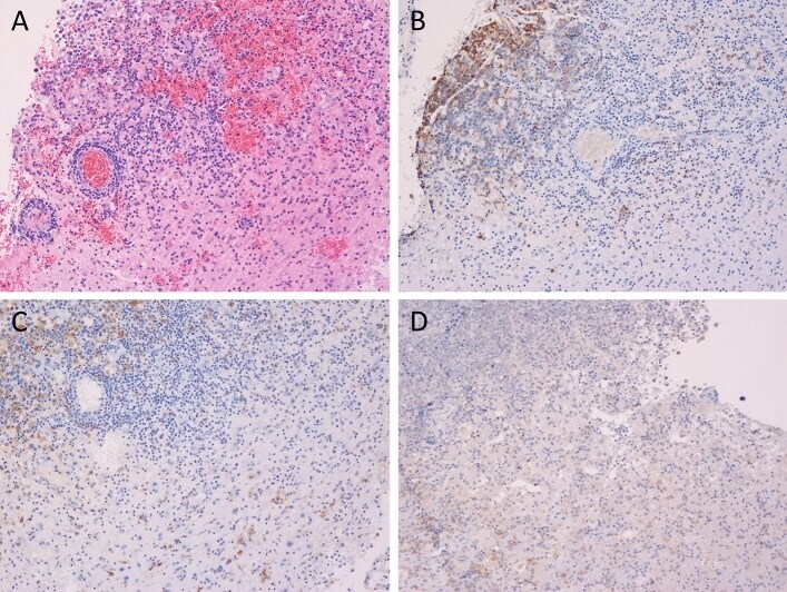
- Figure 3 The histopathological findings in the tumoral and peritumoral tissue. Hematoxylin and eosin staining showed the high cellularity, diffuse growth, and perivascular spread of the tumor cells ( A ). The tumor cells were positive for CD20 ( B ). CD68 staining revealed the existence of intratumoral macrophages and peritumoral macrophages ( C ). The macrophages were positive for Siglec-15 while the tumor cells were negative for Siglec-15 ( D ). (magnification x 100). Dark red-brown color represented the presence of antigens in ( B - D ).
- Submitted by
- Invitrogen Antibodies (provider)
- Main image
- Experimental details
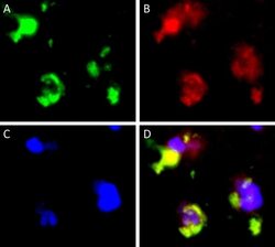
- Figure 4 Double-immunofluorescent staining of CD68 and Siglec-15. CD68 staining ( A green). Siglec-15 staining ( B red). DAPI staining ( C blue). Merged images ( D ). Representative images of PCNSL peritumoral tissue with the co-expression of CD68 and Siglec-15. (magnification x 1000).
