 Explore
 Validate
 Learn
Learn Immunohistochemistry
Antibody data
- Antibody Data
- Antigen structure
- References [2]
- Comments [0]
- Validations
- Immunohistochemistry [1]
Submit
Validation data
Reference
Comment
Report error
- Product number
- MAB20501 - Provider product page

- Provider
- R&D Systems
- Product name
- Human MIA Antibody
- Antibody type
- Monoclonal
- Description
- Protein A or G purified from hybridoma culture supernatant. Detects human MIA in direct ELISAs and Western blots. In direct ELISAs and Western blots, no cross-reactivity with recombinant mouse IL-5 or recombinant rat IL-5 is observed.
- Reactivity
- Human
- Host
- Mouse
- Conjugate
- Unconjugated
- Antigen sequence
Q16674- Isotype
- IgG
- Antibody clone number
- 294203
- Vial size
- 100 ug
- Concentration
- LYOPH
- Storage
- Use a manual defrost freezer and avoid repeated freeze-thaw cycles. 12 months from date of receipt, -20 to -70 °C as supplied. 1 month, 2 to 8 °C under sterile conditions after reconstitution. 6 months, -20 to -70 °C under sterile conditions after reconstitution.
Submitted references Storkhead box 2 and melanoma inhibitory activity promote oral squamous cell carcinoma progression.
MIA is a potential biomarker for tumour load in neurofibromatosis type 1.
Sasahira T, Nishiguchi Y, Fujiwara R, Kurihara M, Kirita T, Bosserhoff AK, Kuniyasu H
Oncotarget 2016 May 3;7(18):26751-64
Oncotarget 2016 May 3;7(18):26751-64
MIA is a potential biomarker for tumour load in neurofibromatosis type 1.
Kolanczyk M, Mautner V, Kossler N, Nguyen R, Kühnisch J, Zemojtel T, Jamsheer A, Wegener E, Thurisch B, Tinschert S, Holtkamp N, Park SJ, Birch P, Kendler D, Harder A, Mundlos S, Kluwe L
BMC medicine 2011 Jul 4;9:82
BMC medicine 2011 Jul 4;9:82
No comments: Submit comment
Supportive validation
- Submitted by
- R&D Systems (provider)
- Main image
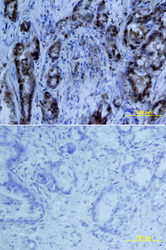
- Experimental details
- MIA in Human Pancreas. MIA was detected in immersion fixed paraffin-embedded sections of human pancreas array using Mouse Anti-Human MIA Monoclonal Antibody (Catalog # MAB20501) at 25 µg/mL overnight at 4 °C. Tissue was stained using the Anti-Mouse HRP-DAB Cell & Tissue Staining Kit (brown; Catalog # CTS002) and counterstained with hematoxylin (blue). Lower panel shows a lack of labeling if primary antibodies are omitted and tissue is stained only with secondary antibody followed by incubation with detection reagents. View our protocol for Chromogenic IHC Staining of Paraffin-embedded Tissue Sections.
