 Explore
 Validate
 Learn
Learn Western blot
 Immunocytochemistry
 Immunohistochemistry
Antibody data
- Antibody Data
- Antigen structure
- References [7]
- Comments [0]
- Validations
- Western blot [1]
- Immunocytochemistry [1]
Submit
Validation data
Reference
Comment
Report error
- Product number
- HPA001031 - Provider product page

- Provider
- Atlas Antibodies
- Proper citation
- Atlas Antibodies Cat#HPA001031, RRID:AB_1078610
- Product name
- Anti-CSTA
- Antibody type
- Polyclonal
- Description
- Polyclonal Antibody against Human CSTA, Gene description: cystatin A (stefin A), Alternative Gene Names: STF1, STFA, Validated applications: WB, IHC, ICC, Uniprot ID: P01040, Storage: Store at +4°C for short term storage. Long time storage is recommended at -20°C.
- Reactivity
- Human
- Host
- Rabbit
- Conjugate
- Unconjugated
- Isotype
- IgG
- Vial size
- 100 µl
- Concentration
- 0.1 mg/ml
- Storage
- Store at +4°C for short term storage. Long time storage is recommended at -20°C.
- Handling
- The antibody solution should be gently mixed before use.
Submitted references Opposing Wnt signals regulate cervical squamocolumnar homeostasis and emergence of metaplasia
Hepatitis B Virus X Protein (HBx) Suppresses Transcription Factor EB (TFEB) Resulting in Stabilization of Integrin Beta 1 (ITGB1) in Hepatocellular Carcinoma Cells
A High-throughput Bead-based Affinity Assay Enables Analysis of Genital Protein Signatures in Women At Risk of HIV Infection
Clinicopathological significance of cystatin A expression in progression of esophageal squamous cell carcinoma
Particular gene upregulation and p53 heterogeneous expression in TP53-mutated maxillary carcinoma.
Expression profiling of microdissected cell populations selected from basal cells in normal epidermis and basal cell carcinoma
From Gene Expression Analysis to Tissue Microarrays
Chumduri C, Gurumurthy R, Berger H, Dietrich O, Kumar N, Koster S, Brinkmann V, Hoffmann K, Drabkina M, Arampatzi P, Son D, Klemm U, Mollenkopf H, Herbst H, Mangler M, Vogel J, Saliba A, Meyer T
Nature Cell Biology 2021;23(2):184-197
Nature Cell Biology 2021;23(2):184-197
Hepatitis B Virus X Protein (HBx) Suppresses Transcription Factor EB (TFEB) Resulting in Stabilization of Integrin Beta 1 (ITGB1) in Hepatocellular Carcinoma Cells
Zhang C, Yang H, Pan L, Zhao G, Zhang R, Zhang T, Xiao Z, Tong Y, Zhang Y, Hu R, Pandol S, Han Y
Cancers 2021;13(5):1181
Cancers 2021;13(5):1181
A High-throughput Bead-based Affinity Assay Enables Analysis of Genital Protein Signatures in Women At Risk of HIV Infection
Månberg A, Bradley F, Qundos U, Guthrie B, Birse K, Noël-Romas L, Lindskog C, Bosire R, Kiarie J, Farquhar C, Burgener A, Nilsson P, Broliden K
Molecular & Cellular Proteomics 2019;18(3):461-476
Molecular & Cellular Proteomics 2019;18(3):461-476
Clinicopathological significance of cystatin A expression in progression of esophageal squamous cell carcinoma
Shiba D, Terayama M, Yamada K, Hagiwara T, Oyama C, Tamura-Nakano M, Igari T, Yokoi C, Soma D, Nohara K, Yamashita S, Dohi T, Kawamura Y
Medicine 2018;97(15):e0357
Medicine 2018;97(15):e0357
Particular gene upregulation and p53 heterogeneous expression in TP53-mutated maxillary carcinoma.
Kudo I, Esumi M, Kusumi Y, Furusaka T, Oshima T
Oncology letters 2017 Oct;14(4):4633-4640
Oncology letters 2017 Oct;14(4):4633-4640
Expression profiling of microdissected cell populations selected from basal cells in normal epidermis and basal cell carcinoma
Asplund A, Gry Björklund M, Sundquist C, Strömberg S, Edlund K, Östman A, Nilsson P, Pontén F, Lundeberg J
British Journal of Dermatology 2008;158(3):527-538
British Journal of Dermatology 2008;158(3):527-538
From Gene Expression Analysis to Tissue Microarrays
Ek S, Andréasson U, Hober S, Kampf C, Pontén F, Uhlén M, Merz H, Borrebaeck C
Molecular & Cellular Proteomics 2006;5(6):1072-1081
Molecular & Cellular Proteomics 2006;5(6):1072-1081
No comments: Submit comment
Enhanced validation
- Submitted by
- Atlas Antibodies (provider)
- Enhanced method
- Genetic validation
- Main image
- Experimental details
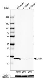
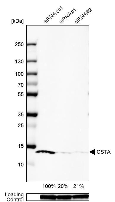
- Western blot analysis in U-87MG ATCC cells transfected with control siRNA, target specific siRNA probe #1 and #2, using Anti-CSTA antibody. Remaining relative intensity is presented. Loading control: Anti-GAPDH.
- Sample type
- Human
- Protocol
- Protocol
Supportive validation
- Submitted by
- Atlas Antibodies (provider)
- Main image
- Experimental details
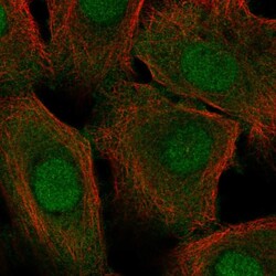
- Immunofluorescent staining of human cell line hTCEpi shows localization to nucleus & cytosol.
- Sample type
- Human
