 Explore
 Validate
 Learn
Learn Western blot
 Immunohistochemistry
Antibody data
- Antibody Data
- Antigen structure
- References [5]
- Comments [0]
- Validations
- Immunohistochemistry [1]
Submit
Validation data
Reference
Comment
Report error
- Product number
- HPA000980 - Provider product page

- Provider
- Atlas Antibodies
- Proper citation
- Atlas Antibodies Cat#HPA000980, RRID:AB_1078170
- Product name
- Anti-AOC3
- Antibody type
- Polyclonal
- Description
- Polyclonal Antibody against Human AOC3, Gene description: amine oxidase, copper containing 3, Alternative Gene Names: HPAO, VAP-1, VAP1, Validated applications: WB, IHC, Uniprot ID: Q16853, Storage: Store at +4°C for short term storage. Long time storage is recommended at -20°C.
- Reactivity
- Human
- Host
- Rabbit
- Conjugate
- Unconjugated
- Isotype
- IgG
- Vial size
- 100 µl
- Concentration
- 0.05 mg/ml
- Storage
- Store at +4°C for short term storage. Long time storage is recommended at -20°C.
- Handling
- The antibody solution should be gently mixed before use.
Submitted references Exometabolomic Analysis of Decidualizing Human Endometrial Stromal and Perivascular Cells
Vascular Adhesion Protein-1 (VAP-1) as Predictor of Radiographic Severity in Symptomatic Knee Osteoarthritis in the New York University Cohort
Evaluation of serum and tissue levels of VAP-1 in colorectal cancer
Vascular adhesion protein-1 promotes liver inflammation and drives hepatic fibrosis
From Gene Expression Analysis to Tissue Microarrays
Harden S, Zhou J, Gharanei S, Diniz-da-Costa M, Lucas E, Cui L, Murakami K, Fang J, Chen Q, Brosens J, Lee Y
Frontiers in Cell and Developmental Biology 2021;9
Frontiers in Cell and Developmental Biology 2021;9
Vascular Adhesion Protein-1 (VAP-1) as Predictor of Radiographic Severity in Symptomatic Knee Osteoarthritis in the New York University Cohort
Bournazou E, Samuels J, Zhou H, Krasnokutsky S, Patel J, Han T, Bencardino J, Rybak L, Abramson S, Junker U, Brown K, Attur M
International Journal of Molecular Sciences 2019;20(11):2642
International Journal of Molecular Sciences 2019;20(11):2642
Evaluation of serum and tissue levels of VAP-1 in colorectal cancer
Ward S, Weston C, Shepherd E, Hejmadi R, Ismail T, Adams D
BMC Cancer 2016;16(1)
BMC Cancer 2016;16(1)
Vascular adhesion protein-1 promotes liver inflammation and drives hepatic fibrosis
Weston C, Shepherd E, Claridge L, Rantakari P, Curbishley S, Tomlinson J, Hubscher S, Reynolds G, Aalto K, Anstee Q, Jalkanen S, Salmi M, Smith D, Day C, Adams D
Journal of Clinical Investigation 2014;125(2):501-520
Journal of Clinical Investigation 2014;125(2):501-520
From Gene Expression Analysis to Tissue Microarrays
Ek S, Andréasson U, Hober S, Kampf C, Pontén F, Uhlén M, Merz H, Borrebaeck C
Molecular & Cellular Proteomics 2006;5(6):1072-1081
Molecular & Cellular Proteomics 2006;5(6):1072-1081
No comments: Submit comment
Supportive validation
- Submitted by
- Atlas Antibodies (provider)
- Enhanced method
- Orthogonal validation
- Main image
- Experimental details
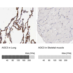
- Immunohistochemistry analysis in human lung and skeletal muscle tissues using HPA000980 antibody. Corresponding AOC3 RNA-seq data are presented for the same tissues.
- Sample type
- Human
- Protocol
- Protocol
