Explore
 Validate
 Learn
Learn Western blot
Antibody data
- Antibody Data
- Antigen structure
- References [2]
- Comments [0]
- Validations
- Western blot [1]
- Immunohistochemistry [6]
Submit
Validation data
Reference
Comment
Report error
- Product number
- NBP1-89671 - Provider product page

- Provider
- Novus Biologicals
- Proper citation
- Novus Cat#NBP1-89671, RRID:AB_11015219
- Product name
- Rabbit Polyclonal VAP-1/AOC3 Antibody
- Antibody type
- Polyclonal
- Description
- Immunogen affinity purified. Specificity of human VAP-1/AOC3 antibody verified on a Protein Array containing target protein plus 383 other non-specific proteins.
- Reactivity
- Human
- Host
- Rabbit
- Isotype
- IgG
- Vial size
- 0.1 ml
- Storage
- Store at 4C short term. Aliquot and store at -20C long term. Avoid freeze-thaw cycles.
Submitted references Vascular adhesion protein-1 promotes liver inflammation and drives hepatic fibrosis.
From gene expression analysis to tissue microarrays: a rational approach to identify therapeutic and diagnostic targets in lymphoid malignancies.
Weston CJ, Shepherd EL, Claridge LC, Rantakari P, Curbishley SM, Tomlinson JW, Hubscher SG, Reynolds GM, Aalto K, Anstee QM, Jalkanen S, Salmi M, Smith DJ, Day CP, Adams DH
The Journal of clinical investigation 2015 Feb;125(2):501-20
The Journal of clinical investigation 2015 Feb;125(2):501-20
From gene expression analysis to tissue microarrays: a rational approach to identify therapeutic and diagnostic targets in lymphoid malignancies.
Ek S, Andréasson U, Hober S, Kampf C, Pontén F, Uhlén M, Merz H, Borrebaeck CA
Molecular & cellular proteomics : MCP 2006 Jun;5(6):1072-81
Molecular & cellular proteomics : MCP 2006 Jun;5(6):1072-81
No comments: Submit comment
Supportive validation
- Submitted by
- Novus Biologicals (provider)
- Main image

- Experimental details
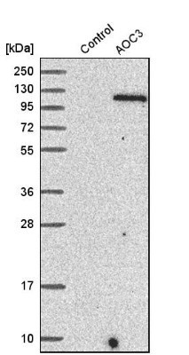
- Western Blot: VAP-1/AOC3 Antibody [NBP1-89671] - Analysis in control (vector only transfected HEK293T lysate) and aOC3 over-expression lysate (Co-expressed with a C-terminal myc-DDK tag (3.1 kDa) in mammalian HEK293T cells).
Supportive validation
- Submitted by
- Novus Biologicals (provider)
- Main image
- Experimental details
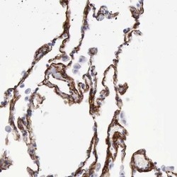
- Immunohistochemistry-Paraffin: VAP-1/AOC3 Antibody [NBP1-89671] - Staining of human lung shows high expression.

- Submitted by
- Novus Biologicals (provider)
- Main image
- Experimental details
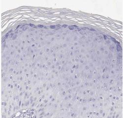
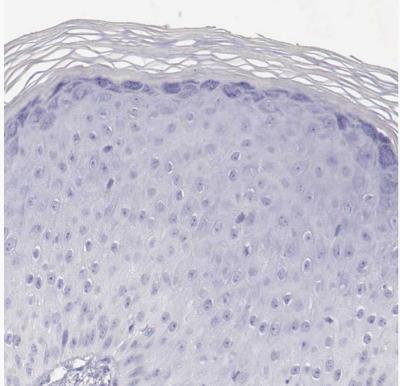
- Immunohistochemistry-Paraffin: VAP-1/AOC3 Antibody [NBP1-89671] - Staining of human skin shows no positivity in squamous epithelial cells as expected.
- Submitted by
- Novus Biologicals (provider)
- Main image
- Experimental details
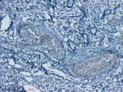
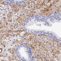
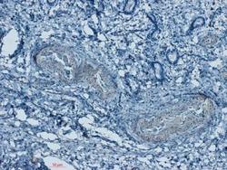
- Immunohistochemistry-Paraffin: VAP-1/AOC3 Antibody [NBP1-89671] - Staining of human prostate shows moderate membranous positivity in smooth muscle cells.

- Submitted by
- Novus Biologicals (provider)
- Main image
- Experimental details
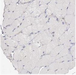
- Immunohistochemistry-Paraffin: VAP-1/AOC3 Antibody [NBP1-89671] - Staining of human skeletal muscle shows no positivity in myocytes as expected.
- Submitted by
- Novus Biologicals (provider)
- Main image
- Experimental details
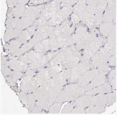
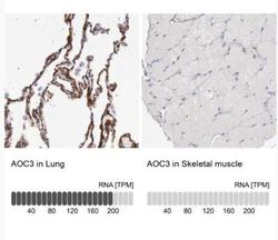
- Immunohistochemistry-Paraffin: VAP-1/AOC3 Antibody [NBP1-89671] - Analysis in human lung and skeletal muscle tissues. Corresponding AOC3 RNA-seq data are presented for the same tissues.

- Submitted by
- Novus Biologicals (provider)
- Main image
- Experimental details
- Immunohistochemistry-Paraffin: VAP-1/AOC3 Antibody [NBP1-89671] - Equine endometrium. All vascular smooth muscle was positively stained. IHC-P image submitted by a verified customer review.