Explore
 Validate
 Learn
Learn Western blot
 ELISA
ELISAAntibody data
- Antibody Data
- Antigen structure
- References [0]
- Comments [0]
- Validations
- Western blot [2]
- Immunohistochemistry [1]
Submit
Validation data
Reference
Comment
Report error
- Product number
- LS-C744821 - Provider product page

- Provider
- LSBio
- Product name
- PDCD4 Antibody (phospho-Ser457, clone 9G6) LS-C744821
- Antibody type
- Monoclonal
- Description
- Protein A affinity chromatography
- Reactivity
- Human, Mouse, Rat, Xenopus
- Host
- Mouse
- Isotype
- IgG
- Antibody clone number
- 9G6
- Storage
- Store vial at -20°C or below prior to opening. Dilute 1:10 to minimize loss. Store the vial at -20°C or below after dilution. Avoid freeze-thaw cycles.
No comments: Submit comment
Supportive validation
- Submitted by
- LSBio (provider)
- Enhanced method
- Genetic validation
- Main image

- Experimental details
- Western blot using the Protein A purified Mouse Monoclonal anti-Pdcd4 pS457 antibody shows detection of phosphorylated Pdcd4 (indicated by arrowhead at ~62 kDa) in NIH-3T3 cells after 5 min treatment with 30 ng/mL PDGF (lane 2). No reactivity is seen for unstimulated (non-phosphorylated) NIH 3T3 cells (lane 1). The membrane was probed with the primary antibody at a 1:2,000 dilution, overnight at 4° C. For detection HRP conjugated Rb-a-Mouse IgG was used at a 1:20,000 dilution in blocking buffer for 1 h at 4° C followed by visualization using a Biospectrum imaging system (UVP).

- Submitted by
- LSBio (provider)
- Enhanced method
- Genetic validation
- Main image

- Experimental details
- Western blot using Protein A purified Mouse Monoclonal anti-Pdcd4 pS457 antibody against recombinant PDCD4 protein. Membrane was blocked in 1% BSA-TBS-T for 30 min RT and probed with 1° Ab Ms-A-Pdcd4pS457 1:1000 (o/n 4°C in 1% BSA-TBS-T) followed by 2° Ab Peroxidase Conjugated Rabbit anti Ms CUST10M at 1:40,000 in MB-070 30 min RT. Bands at ~62 kD and ~32 kD were detected.

Supportive validation
- Submitted by
- LSBio (provider)
- Main image

- Experimental details
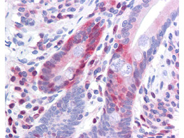
- Antibody 200-301-964 has been tested in immunohistochemistry, analyzed by an anatomic pathologist and validated for use in IHC applications against formalin-fixed, paraffin-embedded human tissues. The antibody was serially diluted and tested at a range of concentrations on at least 22 different human formalin-fixed, paraffin archival tissues, and positive and negative tissues were scored and compared to the published literature on the expression and function of the gene. A representative image from positively stained small intestine shows the localization of the anti Pdcd4 antibody as the precipitated red signal, with a hematoxylin purple nuclear counterstain.