Explore
 Validate
 Learn
Learn Western blot
Antibody data
- Antibody Data
- Antigen structure
- References [7]
- Comments [0]
- Validations
- Western blot [2]
- Immunohistochemistry [7]
Submit
Validation data
Reference
Comment
Report error
- Product number
- NBP1-83302 - Provider product page

- Provider
- Novus Biologicals
- Proper citation
- Novus Cat#NBP1-83302, RRID:AB_11005007
- Product name
- Rabbit Polyclonal PDCD4 Antibody
- Antibody type
- Polyclonal
- Description
- Immunogen affinity purified. Specificity of human, mouse, rat PDCD4 antibody verified on a Protein Array containing target protein plus 383 other non-specific proteins.
- Reactivity
- Human, Mouse, Rat
- Host
- Rabbit
- Isotype
- IgG
- Vial size
- 0.1 ml
- Storage
- Store at 4C short term. Aliquot and store at -20C long term. Avoid freeze-thaw cycles.
Submitted references MicroRNA profiles in familial and sporadic medullary thyroid carcinoma: preliminary relationships with RET status and outcome.
Programmed cell death 4 nuclear loss and miR-21 or activated Akt overexpression in esophageal squamous cell carcinogenesis.
PDCD4 nuclear loss inversely correlates with miR-21 levels in colon carcinogenesis.
Programmed cell death 4 (PDCD4) expression during multistep Barrett's carcinogenesis.
MicroRNA expression profiling of human metastatic cancers identifies cancer gene targets.
Tissue profiling of the mammalian central nervous system using human antibody-based proteomics.
From gene expression analysis to tissue microarrays: a rational approach to identify therapeutic and diagnostic targets in lymphoid malignancies.
Mian C, Pennelli G, Fassan M, Balistreri M, Barollo S, Cavedon E, Galuppini F, Pizzi M, Vianello F, Pelizzo MR, Girelli ME, Rugge M, Opocher G
Thyroid : official journal of the American Thyroid Association 2012 Sep;22(9):890-6
Thyroid : official journal of the American Thyroid Association 2012 Sep;22(9):890-6
Programmed cell death 4 nuclear loss and miR-21 or activated Akt overexpression in esophageal squamous cell carcinogenesis.
Fassan M, Realdon S, Pizzi M, Balistreri M, Battaglia G, Zaninotto G, Ancona E, Rugge M
Diseases of the esophagus : official journal of the International Society for Diseases of the Esophagus 2012 Apr;25(3):263-8
Diseases of the esophagus : official journal of the International Society for Diseases of the Esophagus 2012 Apr;25(3):263-8
PDCD4 nuclear loss inversely correlates with miR-21 levels in colon carcinogenesis.
Fassan M, Pizzi M, Giacomelli L, Mescoli C, Ludwig K, Pucciarelli S, Rugge M
Virchows Archiv : an international journal of pathology 2011 Apr;458(4):413-9
Virchows Archiv : an international journal of pathology 2011 Apr;458(4):413-9
Programmed cell death 4 (PDCD4) expression during multistep Barrett's carcinogenesis.
Fassan M, Pizzi M, Battaglia G, Giacomelli L, Parente P, Bocus P, Ancona E, Rugge M
Journal of clinical pathology 2010 Aug;63(8):692-6
Journal of clinical pathology 2010 Aug;63(8):692-6
MicroRNA expression profiling of human metastatic cancers identifies cancer gene targets.
Baffa R, Fassan M, Volinia S, O'Hara B, Liu CG, Palazzo JP, Gardiman M, Rugge M, Gomella LG, Croce CM, Rosenberg A
The Journal of pathology 2009 Oct;219(2):214-21
The Journal of pathology 2009 Oct;219(2):214-21
Tissue profiling of the mammalian central nervous system using human antibody-based proteomics.
Mulder J, Björling E, Jonasson K, Wernérus H, Hober S, Hökfelt T, Uhlén M
Molecular & cellular proteomics : MCP 2009 Jul;8(7):1612-22
Molecular & cellular proteomics : MCP 2009 Jul;8(7):1612-22
From gene expression analysis to tissue microarrays: a rational approach to identify therapeutic and diagnostic targets in lymphoid malignancies.
Ek S, Andréasson U, Hober S, Kampf C, Pontén F, Uhlén M, Merz H, Borrebaeck CA
Molecular & cellular proteomics : MCP 2006 Jun;5(6):1072-81
Molecular & cellular proteomics : MCP 2006 Jun;5(6):1072-81
No comments: Submit comment
Supportive validation
- Submitted by
- Novus Biologicals (provider)
- Main image

- Experimental details
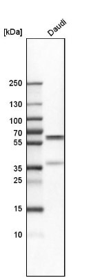
- Western Blot: PDCD4 Antibody [NBP1-83302] - Analysis in human cell line Daudi.
- Submitted by
- Novus Biologicals (provider)
- Main image

- Experimental details
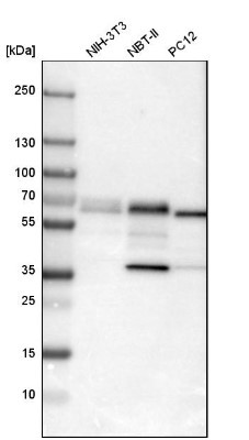
- Western Blot: PDCD4 Antibody [NBP1-83302] - Western blot analysis in mouse cell line NIH-3T3, rat cell line NBT-II and rat cell line pC12.
Supportive validation
- Submitted by
- Novus Biologicals (provider)
- Main image

- Experimental details
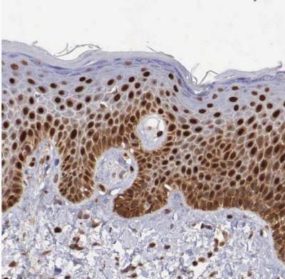
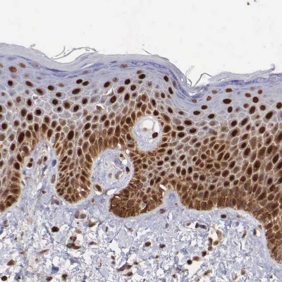
- Immunohistochemistry-Paraffin: PDCD4 Antibody [NBP1-83302] - Staining of human skin shows strong nuclear positivity in epidermal cells.
- Submitted by
- Novus Biologicals (provider)
- Main image

- Experimental details
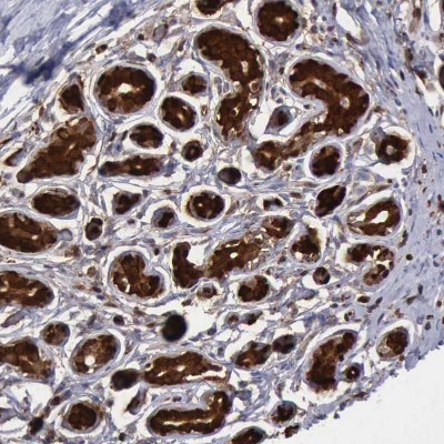
- Immunohistochemistry-Paraffin: PDCD4 Antibody [NBP1-83302] - Staining of human breast shows strong nuclear positivity in glandular cells.
- Submitted by
- Novus Biologicals (provider)
- Main image

- Experimental details
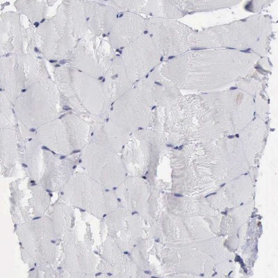
- Immunohistochemistry-Paraffin: PDCD4 Antibody [NBP1-83302] - Staining of human skeletal muscle shows no positivity in myocytes as expected.
- Submitted by
- Novus Biologicals (provider)
- Main image

- Experimental details
- Immunohistochemistry-Paraffin: PDCD4 Antibody [NBP1-83302] - Staining of human skin shows strong nuclear positivity in epidermal cells.
- Submitted by
- Novus Biologicals (provider)
- Main image

- Experimental details
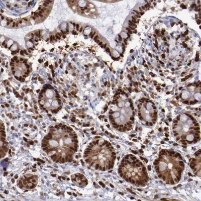
- Immunohistochemistry-Paraffin: PDCD4 Antibody [NBP1-83302] - Staining of human small intestine shows strong nuclear positivity in glandular cells.
- Submitted by
- Novus Biologicals (provider)
- Main image

- Experimental details
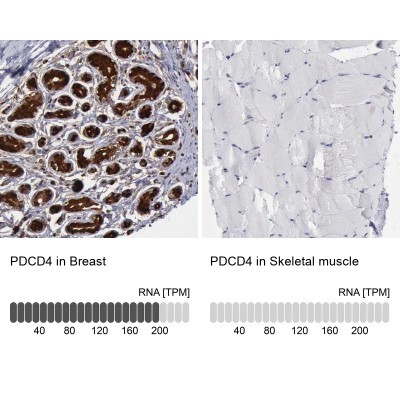
- Immunohistochemistry-Paraffin: PDCD4 Antibody [NBP1-83302] - Staining in human breast and skeletal muscle tissues . Corresponding PDCD4 RNA-seq data are presented for the same tissues.
- Submitted by
- Novus Biologicals (provider)
- Main image

- Experimental details
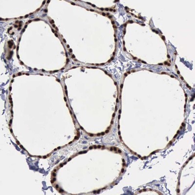
- Immunohistochemistry-Paraffin: PDCD4 Antibody [NBP1-83302] - Staining of human thyroid gland shows strong nuclear positivity in glandular cells.