Explore
 Validate
 Learn
Learn Western blot
 Immunohistochemistry
Antibody data
- Antibody Data
- Antigen structure
- References [9]
- Comments [0]
- Validations
- Immunohistochemistry [1]
Submit
Validation data
Reference
Comment
Report error
- Product number
- HPA001032 - Provider product page

- Provider
- Atlas Antibodies
- Proper citation
- Atlas Antibodies Cat#HPA001032, RRID:AB_1079586
- Product name
- Anti-PDCD4
- Antibody type
- Polyclonal
- Description
- Polyclonal Antibody against Human PDCD4, Gene description: programmed cell death 4 (neoplastic transformation inhibitor), Alternative Gene Names: H731, Validated applications: IHC, WB, Uniprot ID: Q53EL6, Storage: Store at +4°C for short term storage. Long time storage is recommended at -20°C.
- Reactivity
- Human, Mouse, Rat
- Host
- Rabbit
- Conjugate
- Unconjugated
- Isotype
- IgG
- Vial size
- 100 µl
- Concentration
- 0.1 mg/ml
- Storage
- Store at +4°C for short term storage. Long time storage is recommended at -20°C.
- Handling
- The antibody solution should be gently mixed before use.
Submitted references Programmed cell death 4 (PDCD4) as a novel prognostic marker for papillary thyroid carcinoma.
MiR-21 over-expression and Programmed Cell Death 4 down-regulation features malignant pleural mesothelioma.
Programmed cell death 4 nuclear loss and miR-21 or activated Akt overexpression in esophageal squamous cell carcinogenesis
MicroRNA profiles in familial and sporadic medullary thyroid carcinoma: preliminary relationships with RET status and outcome.
PDCD4 nuclear loss inversely correlates with miR-21 levels in colon carcinogenesis
Programmed cell death 4 (PDCD4) expression during multistep Barrett's carcinogenesis.
MicroRNA expression profiling of human metastatic cancers identifies cancer gene targets
Tissue profiling of the mammalian central nervous system using human antibody-based proteomics.
From Gene Expression Analysis to Tissue Microarrays
Galuppini F, Fassan M, Bertazza L, Barollo S, Cascione L, Watutantrige-Fernando S, Lazzarin V, Simonato P, Vianello F, Rugge M, Mian C, Pennelli G
Cancer management and research 2019;11:7845-7855
Cancer management and research 2019;11:7845-7855
MiR-21 over-expression and Programmed Cell Death 4 down-regulation features malignant pleural mesothelioma.
Nicolè L, Cappellesso R, Sanavia T, Guzzardo V, Fassina A
Oncotarget 2018 Apr 3;9(25):17300-17308
Oncotarget 2018 Apr 3;9(25):17300-17308
Programmed cell death 4 nuclear loss and miR-21 or activated Akt overexpression in esophageal squamous cell carcinogenesis
Fassan M, Realdon S, Pizzi M, Balistreri M, Battaglia G, Zaninotto G, Ancona E, Rugge M
Diseases of the Esophagus 2012;25(3):263-268
Diseases of the Esophagus 2012;25(3):263-268
MicroRNA profiles in familial and sporadic medullary thyroid carcinoma: preliminary relationships with RET status and outcome.
Mian C, Pennelli G, Fassan M, Balistreri M, Barollo S, Cavedon E, Galuppini F, Pizzi M, Vianello F, Pelizzo MR, Girelli ME, Rugge M, Opocher G
Thyroid : official journal of the American Thyroid Association 2012 Sep;22(9):890-6
Thyroid : official journal of the American Thyroid Association 2012 Sep;22(9):890-6
PDCD4 nuclear loss inversely correlates with miR-21 levels in colon carcinogenesis
Fassan M, Pizzi M, Giacomelli L, Mescoli C, Ludwig K, Pucciarelli S, Rugge M
Virchows Archiv 2011;458(4):413-419
Virchows Archiv 2011;458(4):413-419
Programmed cell death 4 (PDCD4) expression during multistep Barrett's carcinogenesis.
Fassan M, Pizzi M, Battaglia G, Giacomelli L, Parente P, Bocus P, Ancona E, Rugge M
Journal of clinical pathology 2010 Aug;63(8):692-6
Journal of clinical pathology 2010 Aug;63(8):692-6
MicroRNA expression profiling of human metastatic cancers identifies cancer gene targets
Baffa R, Fassan M, Volinia S, O'Hara B, Liu C, Palazzo J, Gardiman M, Rugge M, Gomella L, Croce C, Rosenberg A
The Journal of Pathology 2009;219(2):214-221
The Journal of Pathology 2009;219(2):214-221
Tissue profiling of the mammalian central nervous system using human antibody-based proteomics.
Mulder J, Björling E, Jonasson K, Wernérus H, Hober S, Hökfelt T, Uhlén M
Molecular & cellular proteomics : MCP 2009 Jul;8(7):1612-22
Molecular & cellular proteomics : MCP 2009 Jul;8(7):1612-22
From Gene Expression Analysis to Tissue Microarrays
Ek S, Andréasson U, Hober S, Kampf C, Pontén F, Uhlén M, Merz H, Borrebaeck C
Molecular & Cellular Proteomics 2006;5(6):1072-1081
Molecular & Cellular Proteomics 2006;5(6):1072-1081
No comments: Submit comment
Supportive validation
- Submitted by
- Atlas Antibodies (provider)
- Enhanced method
- Orthogonal validation
- Main image
- Experimental details
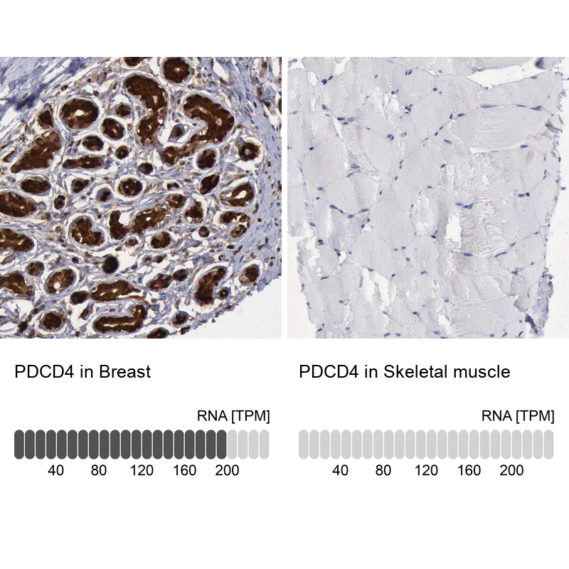
- Immunohistochemistry analysis in human breast and skeletal muscle tissues using HPA001032 antibody. Corresponding PDCD4 RNA-seq data are presented for the same tissues.
- Sample type
- Human
- Protocol
- Protocol