Explore
 Validate
 Learn
Learn Western blot
 ELISA
ELISAAntibody data
- Antibody Data
- Antigen structure
- References [0]
- Comments [0]
- Validations
- Western blot [1]
- Immunohistochemistry [1]
Submit
Validation data
Reference
Comment
Report error
- Product number
- AM08441PU-S - Provider product page

- Provider
- Acris Antibodies GmbH
- Proper citation
- Acris Antibodies GmbH Cat#AM08441PU-S, RRID:AB_2035837
- Product name
- anti PDCD4 pSer457
- Antibody type
- Monoclonal
- Antigen
- Synthetic peptide corresponding to residues surrounding Ser457 of the Human Pdcd4 protein.
- Reactivity
- Human, Mouse, Rat, Xenopus
- Host
- Mouse
- Isotype
- IgG
- Antibody clone number
- 9G6
- Vial size
- 25 µl
- Concentration
- 1.0 mg/ml (by UV absorbance at 280 nm)
No comments: Submit comment
Supportive validation
- Submitted by
- Acris Antibodies GmbH (provider)
- Main image
- Experimental details
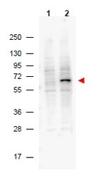
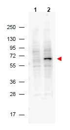
- Western blot Protein A purified Mouse Monoclonal anti-Pdcd4 pS457 antibody shows detection of phosphorylated Pdcd4 (indicated by arrowhead at ~62 kDa) in NIH-3T3 cells after 5 min treatment with 30 ng/mL PDGF (lane 2). No reactivity is seen for unstimulated (non-phosphorylated) NIH 3T3 cells (lane 1). The membrane was probed with the primary antibody at a 1:2,000 dilution, overnight at 4° C. For detection HRP conjugated Rb-a-Mouse IgG was used at a 1:20,000 dilution in blocking buffer (p/n MB-070) for 1 h at 4° C followed by visualization using a Biospectrum imaging system (UVP).
Supportive validation
- Submitted by
- Acris Antibodies GmbH (provider)
- Main image

- Experimental details
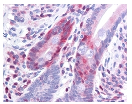
- This antibody has been tested in immunohistochemistry, analyzed by an anatomic pathologist and validated for use in IHC applications against formalin-fixed, paraffin-embedded human tissues. The antibody was serially diluted and tested at a range of concentrations on at least 22 different human formalin-fixed, paraffin archival tissues, and positive and negative tissues were scored and compared to the published literature on the expression and function of the gene. A representative image from positively stained small intestine shows the localization of the anti Pdcd4 antibody as the precipitated red signal, with a hematoxylin purple nuclear counterstain. Image provided courtesy of LifeSpan Biosciences, Seattle, WA