 Explore
 Validate
 Learn
Learn Western blot
 ELISA
ELISAAntibody data
- Antibody Data
- Antigen structure
- References [1]
- Comments [0]
- Validations
- Western blot [1]
- Immunohistochemistry [1]
Submit
Validation data
Reference
Comment
Report error
- Product number
- NB110-60014 - Provider product page

- Provider
- Novus Biologicals
- Proper citation
- Novus Cat#NB110-60014, RRID:AB_905685
- Product name
- Rabbit Polyclonal PDCD4 Antibody
- Antibody type
- Polyclonal
- Description
- Immunogen affinity purified. This antibody is specific for human Pdcd4 protein phosphorylated at Ser457. A BLAST analysis was used to suggest cross-reactivity with Pdcd4 from human, mouse, rat and Xenopus based on 100% identity with the immunizing sequence. Cross-reactivity with Pdcd4 from other sources has not been determined.
- Reactivity
- Human, Mouse, Rat, Xenopus
- Host
- Rabbit
- Isotype
- IgG
- Vial size
- 0.1 mg
- Concentration
- 1 mg/ml
- Storage
- Store at -20C. Avoid freeze-thaw cycles.
Submitted references Up-regulation of miR-21 by HER2/neu signaling promotes cell invasion.
Huang TH, Wu F, Loeb GB, Hsu R, Heidersbach A, Brincat A, Horiuchi D, Lebbink RJ, Mo YY, Goga A, McManus MT
The Journal of biological chemistry 2009 Jul 3;284(27):18515-24
The Journal of biological chemistry 2009 Jul 3;284(27):18515-24
No comments: Submit comment
Supportive validation
- Submitted by
- Novus Biologicals (provider)
- Main image

- Experimental details
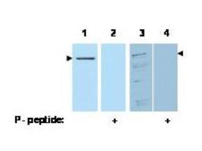
- Western Blot: PDCD4 [p Ser457] Antibody [NB110-60014] - Analysis of Pdcd4 phosphorylated at Ser 457 (arrowheads). Lanes 1 & 2 each contain 100 ng recombinant Pdcd4. Lanes 3 & 4 each contain 30 ug of whole cell extract from 293 HEK cells treated with 20 nM TPA and MG132 proteasome inhibitor for 8 hours. The signal can be competed off with peptide phosphorylated at Ser 457 (Lanes 2 & 4).
Supportive validation
- Submitted by
- Novus Biologicals (provider)
- Main image

- Experimental details
- Immunohistochemistry: PDCD4 [p Ser457] Antibody [NB110-60014] - Used at 1.25 ug/ml to detect signal in a variety of tissues including multi-human, multi-brain and multi-cancer slides. This image shows moderate positive staining of human breast epithelial cells at 40X. Tissue was formalin-fixed and paraffin embedded. The image shows localization of the antibody as the precipitated red signal, with a hematoxylin purple nuclear counterstain.
