Explore
 Validate
 Learn
Learn Western blot
Antibody data
- Antibody Data
- Antigen structure
- References [0]
- Comments [0]
- Validations
- Western blot [2]
- ELISA [1]
- Immunocytochemistry [1]
- Other assay [1]
Submit
Validation data
Reference
Comment
Report error
- Product number
- H00001457-M01 - Provider product page

- Provider
- Invitrogen Antibodies
- Product name
- CSNK2A1 Monoclonal Antibody (3D9)
- Antibody type
- Monoclonal
- Antigen
- Recombinant full-length protein
- Reactivity
- Human
- Host
- Mouse
- Isotype
- IgG
- Antibody clone number
- 3D9
- Vial size
- 100 µg
- Concentration
- See Label
- Storage
- -20° C, Avoid Freeze/Thaw Cycles
No comments: Submit comment
Supportive validation
- Submitted by
- Invitrogen Antibodies (provider)
- Main image
- Experimental details
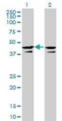
- Western Blot analysis of CSNK2A1 expression in transfected 293T cell line by CSNK2A1 monoclonal antibody (M01), clone 3D9.Lane 1: CSNK2A1 transfected lysate(45.1 KDa).Lane 2: Non-transfected lysate.
- Submitted by
- Invitrogen Antibodies (provider)
- Main image
- Experimental details
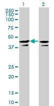
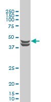
- CSNK2A1 monoclonal antibody (M01), clone 3D9 Western Blot analysis of CSNK2A1 expression in HeLa S3 NE (Cat # L013V3).
Supportive validation
- Submitted by
- Invitrogen Antibodies (provider)
- Main image
- Experimental details
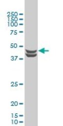
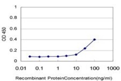
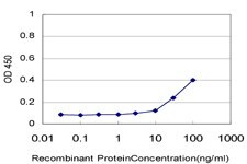
- Detection limit for recombinant GST tagged CSNK2A1 is approximately 3 ng/mL as a capture antibody.
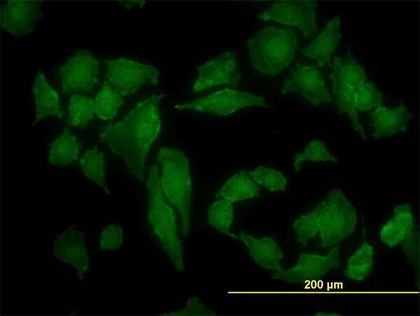
Supportive validation
- Submitted by
- Invitrogen Antibodies (provider)
- Main image
- Experimental details
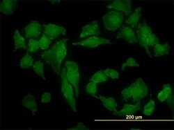
- Immunofluorescence of monoclonal antibody to CSNK2A1 on HeLa cell. Antibody concentration is 10 µg/mL.
Supportive validation
- Submitted by
- Invitrogen Antibodies (provider)
- Main image

- Experimental details
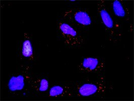
- Proximity Ligation Analysis of protein-protein interactions between TP53 and CSNK2A1. HeLa cells were stained with anti-TP53 rabbit purified polyclonal 1:1200 and anti-CSNK2A1 mouse monoclonal antibody 1:50. Each red dot represents the detection of protein-protein interaction complex, and nuclei were counterstained with DAPI (blue).