Explore
 Validate
 Learn
LearnMA5-23919
antibody from Invitrogen Antibodies
Targeting: NLRP3
AGTAVPRL, AII, AVP, C1orf7, CIAS1, CLR1.1, DFNA34, FCAS, FCU, MWS, NALP3, PYPAF1
 Western blot
 Immunocytochemistry
Antibody data
- Antibody Data
- Antigen structure
- References [8]
- Comments [0]
- Validations
- Immunocytochemistry [2]
- Immunohistochemistry [1]
- Flow cytometry [6]
- Other assay [4]
Submit
Validation data
Reference
Comment
Report error
- Product number
- MA5-23919 - Provider product page

- Provider
- Invitrogen Antibodies
- Product name
- NLRP3 Monoclonal Antibody (768319)
- Antibody type
- Monoclonal
- Antigen
- Recombinant full-length protein
- Description
- In direct ELISAs, 100% cross-reactivity with recombinant human NLRP3/NALP3 is observed. Reconstitute in sterile PBS to a final concentration of 0.5 mg/mL.
- Reactivity
- Human, Mouse
- Host
- Rat
- Isotype
- IgG
- Antibody clone number
- 768319
- Vial size
- 100 μg
- Concentration
- 0.5 mg/mL
- Storage
- -20°C, Avoid Freeze/Thaw Cycles
Submitted references Modified Citrus Pectin Alleviates Cerebral Ischemia/Reperfusion Injury by Inhibiting NLRP3 Inflammasome Activation via TLR4/NF-ĸB Signaling Pathway in Microglia.
miR-25-3p ameliorates SAE by targeting the TLR4/NLRP3 axis.
Huaier extract suppresses non-small cell lung cancer progression through activating NLRP3-dependent pyroptosis.
Sauchinone Protects Renal Mesangial Cell Dysfunction against Angiotensin II by Improving Renal Fibrosis and Inflammation.
Hyperactivity of Innate Immunity Triggers Pain via TLR2-IL-33-Mediated Neuroimmune Crosstalk.
Microbial BMAA elicits mitochondrial dysfunction, innate immunity activation, and Alzheimer's disease features in cortical neurons.
Extracellular ASC exacerbated the recurrent ischemic stroke in an NLRP3-dependent manner.
Monitoring Inflammasome Priming and Activation in Response to Candida albicans.
Cui Y, Zhang NN, Wang D, Meng WH, Chen HS
Journal of inflammation research 2022;15:3369-3385
Journal of inflammation research 2022;15:3369-3385
miR-25-3p ameliorates SAE by targeting the TLR4/NLRP3 axis.
Luo XY, Ying JH, Wang QS
Metabolic brain disease 2022 Aug;37(6):1803-1813
Metabolic brain disease 2022 Aug;37(6):1803-1813
Huaier extract suppresses non-small cell lung cancer progression through activating NLRP3-dependent pyroptosis.
Xie J, Zhuan B, Wang H, Wang Y, Wang X, Yuan Q, Yang Z
Anatomical record (Hoboken, N.J. : 2007) 2021 Feb;304(2):291-301
Anatomical record (Hoboken, N.J. : 2007) 2021 Feb;304(2):291-301
Sauchinone Protects Renal Mesangial Cell Dysfunction against Angiotensin II by Improving Renal Fibrosis and Inflammation.
Yoon JJ, Lee HK, Kim HY, Han BH, Lee HS, Lee YJ, Kang DG
International journal of molecular sciences 2020 Sep 23;21(19)
International journal of molecular sciences 2020 Sep 23;21(19)
Hyperactivity of Innate Immunity Triggers Pain via TLR2-IL-33-Mediated Neuroimmune Crosstalk.
Huang J, Gandini MA, Chen L, M'Dahoma S, Stemkowski PL, Chung H, Muruve DA, Zamponi GW
Cell reports 2020 Oct 6;33(1):108233
Cell reports 2020 Oct 6;33(1):108233
Microbial BMAA elicits mitochondrial dysfunction, innate immunity activation, and Alzheimer's disease features in cortical neurons.
Silva DF, Candeias E, Esteves AR, Magalhães JD, Ferreira IL, Nunes-Costa D, Rego AC, Empadinhas N, Cardoso SM
Journal of neuroinflammation 2020 Nov 5;17(1):332
Journal of neuroinflammation 2020 Nov 5;17(1):332
Extracellular ASC exacerbated the recurrent ischemic stroke in an NLRP3-dependent manner.
He XF, Zeng YX, Li G, Feng YK, Wu C, Liang FY, Zhang Y, Lan Y, Xu GQ, Pei Z
Journal of cerebral blood flow and metabolism : official journal of the International Society of Cerebral Blood Flow and Metabolism 2020 May;40(5):1048-1060
Journal of cerebral blood flow and metabolism : official journal of the International Society of Cerebral Blood Flow and Metabolism 2020 May;40(5):1048-1060
Monitoring Inflammasome Priming and Activation in Response to Candida albicans.
Santana DJ, Anderson FM, O'Meara TR
Current protocols in microbiology 2020 Dec;59(1):e124
Current protocols in microbiology 2020 Dec;59(1):e124
No comments: Submit comment
Supportive validation
- Submitted by
- Invitrogen Antibodies (provider)
- Main image

- Experimental details
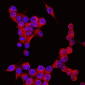
- Immunocytochemistry analysis of NLRP3 in immersion fixed RAW 264.7 mouse monocyte/macrophage cell line. Samples were incubated in NLRP3 monoclonal antibody (Product # MA5-23919) using a dilution of 10 µg/mL for 3 hours at room temperature followed by NorthernLights™ 557-conjugated Anti-Rat IgG Secondary Antibody (red) and counterstained with DAPI (blue). Specific staining was localized to cytoplasm.
- Submitted by
- Invitrogen Antibodies (provider)
- Main image

- Experimental details
- Immunocytochemistry analysis of NLRP3 in immersion fixed RAW 264.7 mouse monocyte/macrophage cell line. Samples were incubated in NLRP3 monoclonal antibody (Product # MA5-23919) using a dilution of 10 µg/mL for 3 hours at room temperature followed by NorthernLights™ 557-conjugated Anti-Rat IgG Secondary Antibody (red) and counterstained with DAPI (blue). Specific staining was localized to cytoplasm.

Supportive validation
- Submitted by
- Invitrogen Antibodies (provider)
- Main image
- Experimental details
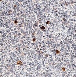
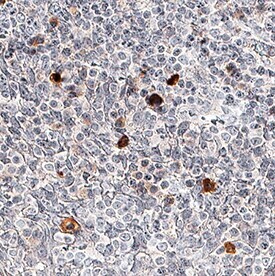
- Immunohistochemical analysis of NLRP3 in immersion fixed paraffin-embedded sections of human tonsil tissue. Samples were incubated in NLRP3 monoclonal antibody (Product # MA5-23919) using a dilution of 5 µg/mL overnight at 4 °C. Before incubation with the primary antibody, tissue was subjected to heat-induced epitope retrieval using Antigen Retrieval Reagent-Basic . Tissue was stained using the Anti-Rat IgG VisUCyte™ HRP Polymer Antibody (brown) and counterstained with hematoxylin (blue). Specific staining was localized to cytoplasm in lymphocytes.
Supportive validation
- Submitted by
- Invitrogen Antibodies (provider)
- Main image
- Experimental details
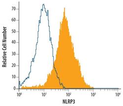
- Flow cytometric analysis of human peripheral blood monocytes was stained with Rat Anti-human/mouse NLRP3/NALP3 Monoclonal Antibody (Product # MA5-23919) or isotype control antibodyopen histogram), followed by Allophycocyanin-conjugated Anti-Rat IgG Secondary Antibody. To facilitate intracellular staining, cells were fixed with paraformaldehyde and permeabilized with saponin.

- Submitted by
- Invitrogen Antibodies (provider)
- Main image

- Experimental details
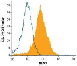
- Flow cytometric analysis of human peripheral blood monocytes was stained with Rat Anti-human/mouse NLRP3/NALP3 Monoclonal Antibody (Product # MA5-23919) or isotype control antibodyopen histogram), followed by Allophycocyanin-conjugated Anti-Rat IgG Secondary Antibody. To facilitate intracellular staining, cells were fixed with paraformaldehyde and permeabilized with saponin.

- Submitted by
- Invitrogen Antibodies (provider)
- Main image
- Experimental details
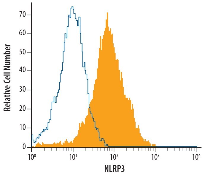
- Flow cytometry of NLRP3 in RAW 264.7 mouse monocyte/macrophage cell line. Samples were incubated in NLRP3 monoclonal antibody (Product # MA5-23919) or isotype control antibody followed by Allophycocyanin-conjugated Anti-Rat IgG Secondary Antibody. To facilitate intracellular staining, cells were fixed with paraformaldehyde and permeabilized with saponin.

- Submitted by
- Invitrogen Antibodies (provider)
- Main image

- Experimental details
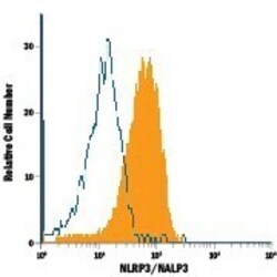
- Flow cytometry of NLRP3 in Human peripheral blood monocytes. Samples were incubated in NLRP3 monoclonal antibody (Product # MA5-23919) or isotype control antibody followed by Allophycocyanin-conjugated Anti-Rat IgG Secondary Antibody. To facilitate intracellular staining, cells were fixed with paraformaldehyde and permeabilized with saponin.

- Submitted by
- Invitrogen Antibodies (provider)
- Main image

- Experimental details
- Flow cytometry of NLRP3 in RAW 264.7 mouse monocyte/macrophage cell line. Samples were incubated in NLRP3 monoclonal antibody (Product # MA5-23919) or isotype control antibody followed by Allophycocyanin-conjugated Anti-Rat IgG Secondary Antibody. To facilitate intracellular staining, cells were fixed with paraformaldehyde and permeabilized with saponin.
- Submitted by
- Invitrogen Antibodies (provider)
- Main image
- Experimental details
- Flow cytometry of NLRP3 in Human peripheral blood monocytes. Samples were incubated in NLRP3 monoclonal antibody (Product # MA5-23919) or isotype control antibody followed by Allophycocyanin-conjugated Anti-Rat IgG Secondary Antibody. To facilitate intracellular staining, cells were fixed with paraformaldehyde and permeabilized with saponin.

Supportive validation
- Submitted by
- Invitrogen Antibodies (provider)
- Main image
- Experimental details
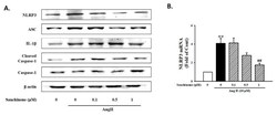
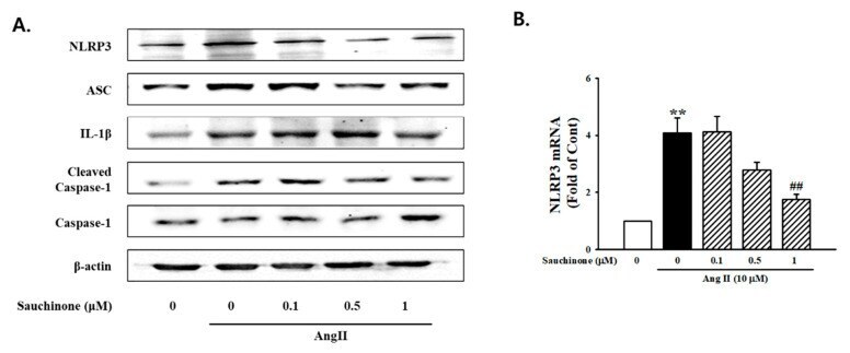
- Figure 5 Inhibition effects of sauchinone on the NOD-like receptor family, pyrin domain-containing-3 (NLRP3) inflammasome in mesangial cells. ( A ) Western blotting and ( B ) real-time PCR showed protein and mRNA levels of NLRP3, ASC, IL-1beta, cleaved caspase-1, and caspase-1 in sauchinone-treated and AngII-stimulated cells at 48 h. beta-actin or GAPDH were used as the internal standard in each sample. ** p < 0.01 vs. control and ## p < 0.01 vs. AngII alone.
- Submitted by
- Invitrogen Antibodies (provider)
- Main image
- Experimental details
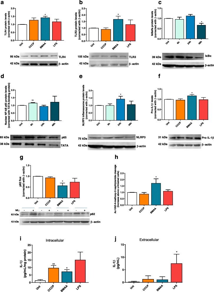
- Fig. 6 BMAA activates neuronal innate immunity. In primary mouse cortical neurons pre-treated with 1 muM CCCP, 3 mM BMAA, and 1 mug/mul LPS for 48 h or with 3 mM BMAA for6, 24 and 48 h, we evaluated a changes in protein levels of TLR4 and b TLR 3 by western blotting. Representative immunoblot and densitometric analysis was performed by the loading of equal amounts of protein corrected with beta-actin. Inflammasome activation was determined by the quantification of c cytosolic IkBalpha protein levels which were determined by western blotting. Representative immunoblot and densitometric analysis was performed by loading equal amounts of protein corrected with beta-actin. d p65 NFkappaB was detected in enriched nuclear fractions by western blotting. Representative immunoblot and densitometric analysis was performed by the loading of equal amounts of protein corrected with TATA-binding protein. e Cytosolic NLRP3 protein levels were evaluated by western blotting. Representative immunoblot and densitometric analysis was performed by loading equal amounts of protein corrected against beta-actin. f Cytosolic pro-IL-1beta protein levels analyzed by western blotting. Representative immunoblot and densitometric analysis was performed by the loading of equal amounts of protein corrected with beta-actin. g p62 flux was evaluated by western blotting after 4-h incubation with or without NH 4 Cl plus leupeptin. Representative immunoblot and densitometric analysis was performed by the loading
- Submitted by
- Invitrogen Antibodies (provider)
- Main image
- Experimental details
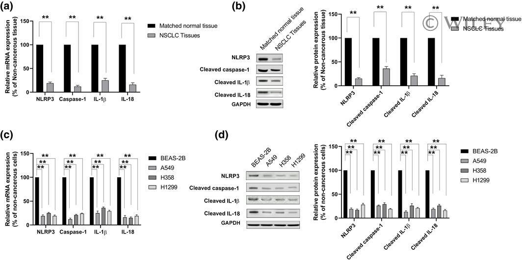
- 1 FIGURE Pyroptosis levels were decreased in non-small cell lung cancer (NSCLC) tissues and cell lines. (a and c) The mRNA levels of pyroptotic-related genes (NLRP3, caspase-1, IL-1beta, and IL-18) in NSCLC tissues, adjacent noncancerous tissues, NSCLC cell lines (A549, H358, and H520), and normal lung epithelial cell line BEAS-2B were analyzed by using qRT-PCR. (b and d) The proteins levels of pyroptotic-related genes (NLRP3, caspase-1, IL-1beta, and IL-18) in NSCLC tissues, adjacent noncancerous tissues, NSCLC cell lines (A549, H358, and H520), and normal lung epithelial cell line BEAS-2B were analyzed by using western blot. GAPDH is chosen as protein controls. ** p < .01
- Submitted by
- Invitrogen Antibodies (provider)
- Main image

- Experimental details
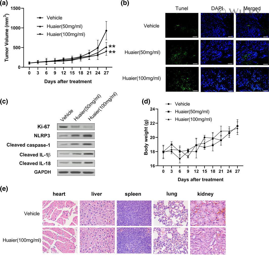
- 6 FIGURE Huaier suppresses tumor growth and induces pyroptosis in vivo. Tumorigenesis assay was performed by subcutaneous injection of H520 cells into flanks of BALB/c nude mice. (a) Tumor volumes were measured at the indicated days post-injection. (b) The effect of huaier on apoptosis in tumor tissues was evaluated by TUNEL assay. (c) The western blot analysis detected the relative expression of Ki-67, NLRP3, caspase-1, IL-1beta, and IL-18 in tumor tissues. (d) Bodyweight of mice in the vehicle group, low dose group (50 mg/ml), and high dose group (100 mg/ml) were evaluated every 3 days. (e) H&E staining was applied to evaluate the changes in the heart, liver, spleen, lung, and kidney tissues. ** p < .01