Explore
 Validate
 Learn
LearnPA5-79740
antibody from Invitrogen Antibodies
Targeting: NLRP3
AGTAVPRL, AII, AVP, C1orf7, CIAS1, CLR1.1, DFNA34, FCAS, FCU, MWS, NALP3, PYPAF1
 Western blot
 Immunocytochemistry
Antibody data
- Antibody Data
- Antigen structure
- References [7]
- Comments [0]
- Validations
- Immunocytochemistry [3]
- Immunohistochemistry [7]
- Flow cytometry [5]
- Other assay [6]
Submit
Validation data
Reference
Comment
Report error
- Product number
- PA5-79740 - Provider product page

- Provider
- Invitrogen Antibodies
- Product name
- NLRP3 Polyclonal Antibody
- Antibody type
- Polyclonal
- Antigen
- Synthetic peptide
- Description
- Reconstitute with 0.2 mL of distilled water to yield a concentration of 500 µg/mL. Positive Control - WB: rat thymus tissue, mouse thyus tissue, mouse RAW264.7 whole cell, mouse J774A.1 whole cell, human THP-1 whole cell. IHC: human colon cancer tissue. Flow: THP-1 cell.
- Reactivity
- Human, Mouse, Rat
- Host
- Rabbit
- Isotype
- IgG
- Vial size
- 100 μg
- Concentration
- 500 μg/mL
- Storage
- -20°C
Submitted references Erythropoietin Enhances Post-ischemic Migration and Phagocytosis and Alleviates the Activation of Inflammasomes in Human Microglial Cells.
Stem Cells From Human Exfoliated Deciduous Teeth Alleviate Liver Cirrhosis via Inhibition of Gasdermin D-Executed Hepatocyte Pyroptosis.
Electroacupuncture Inhibits NLRP3 Activation by Regulating CMPK2 After Spinal Cord Injury.
Anti-Inflammatory Properties of KLS-13019: a Novel GPR55 Antagonist for Dorsal Root Ganglion and Hippocampal Cultures.
Oridonin relieves depressive-like behaviors by inhibiting neuroinflammation and autophagy impairment in rats subjected to chronic unpredictable mild stress.
NU9056, a KAT 5 Inhibitor, Treatment Alleviates Brain Dysfunction by Inhibiting NLRP3 Inflammasome Activation, Affecting Gut Microbiota, and Derived Metabolites in LPS-Treated Mice.
NLRP3 Inflammasome: A Potential Target in Isoflurane Pretreatment Alleviates Stroke-Induced Retinal Injury in Diabetes.
Arik E, Heinisch O, Bienert M, Gubeljak L, Slowik A, Reich A, Schulz JB, Wilhelm T, Huber M, Habib P
Frontiers in cellular neuroscience 2022;16:915348
Frontiers in cellular neuroscience 2022;16:915348
Stem Cells From Human Exfoliated Deciduous Teeth Alleviate Liver Cirrhosis via Inhibition of Gasdermin D-Executed Hepatocyte Pyroptosis.
Chen P, Zhou YK, Han CS, Chen LJ, Wang YM, Zhuang ZM, Lin S, Zhou YH, Jiang JH, Yang RL
Frontiers in immunology 2022;13:860225
Frontiers in immunology 2022;13:860225
Electroacupuncture Inhibits NLRP3 Activation by Regulating CMPK2 After Spinal Cord Injury.
Chen Y, Wu L, Shi M, Zeng D, Hu R, Wu X, Han S, He K, Xu H, Shao X, Ma R
Frontiers in immunology 2022;13:788556
Frontiers in immunology 2022;13:788556
Anti-Inflammatory Properties of KLS-13019: a Novel GPR55 Antagonist for Dorsal Root Ganglion and Hippocampal Cultures.
Brenneman DE, Kinney WA, McDonnell ME, Zhao P, Abood ME, Ward SJ
Journal of molecular neuroscience : MN 2022 Sep;72(9):1859-1874
Journal of molecular neuroscience : MN 2022 Sep;72(9):1859-1874
Oridonin relieves depressive-like behaviors by inhibiting neuroinflammation and autophagy impairment in rats subjected to chronic unpredictable mild stress.
Liang L, Wang H, Hu Y, Bian H, Xiao L, Wang G
Phytotherapy research : PTR 2022 Aug;36(8):3335-3351
Phytotherapy research : PTR 2022 Aug;36(8):3335-3351
NU9056, a KAT 5 Inhibitor, Treatment Alleviates Brain Dysfunction by Inhibiting NLRP3 Inflammasome Activation, Affecting Gut Microbiota, and Derived Metabolites in LPS-Treated Mice.
Chen L, Qing W, Yi Z, Lin G, Peng Q, Zhou F
Frontiers in nutrition 2021;8:701760
Frontiers in nutrition 2021;8:701760
NLRP3 Inflammasome: A Potential Target in Isoflurane Pretreatment Alleviates Stroke-Induced Retinal Injury in Diabetes.
Lin HB, Lin YH, Zhang JY, Guo WJ, Ovcjak A, You ZJ, Feng ZP, Sun HS, Li FX, Zhang HF
Frontiers in cellular neuroscience 2021;15:697449
Frontiers in cellular neuroscience 2021;15:697449
No comments: Submit comment
Supportive validation
- Submitted by
- Invitrogen Antibodies (provider)
- Main image

- Experimental details
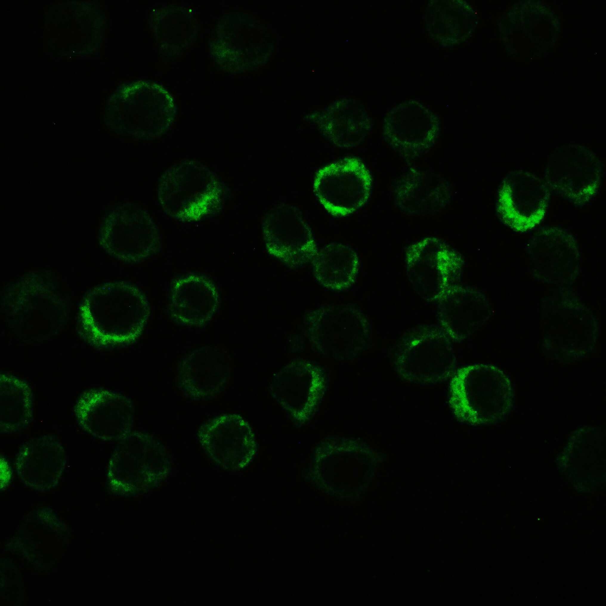
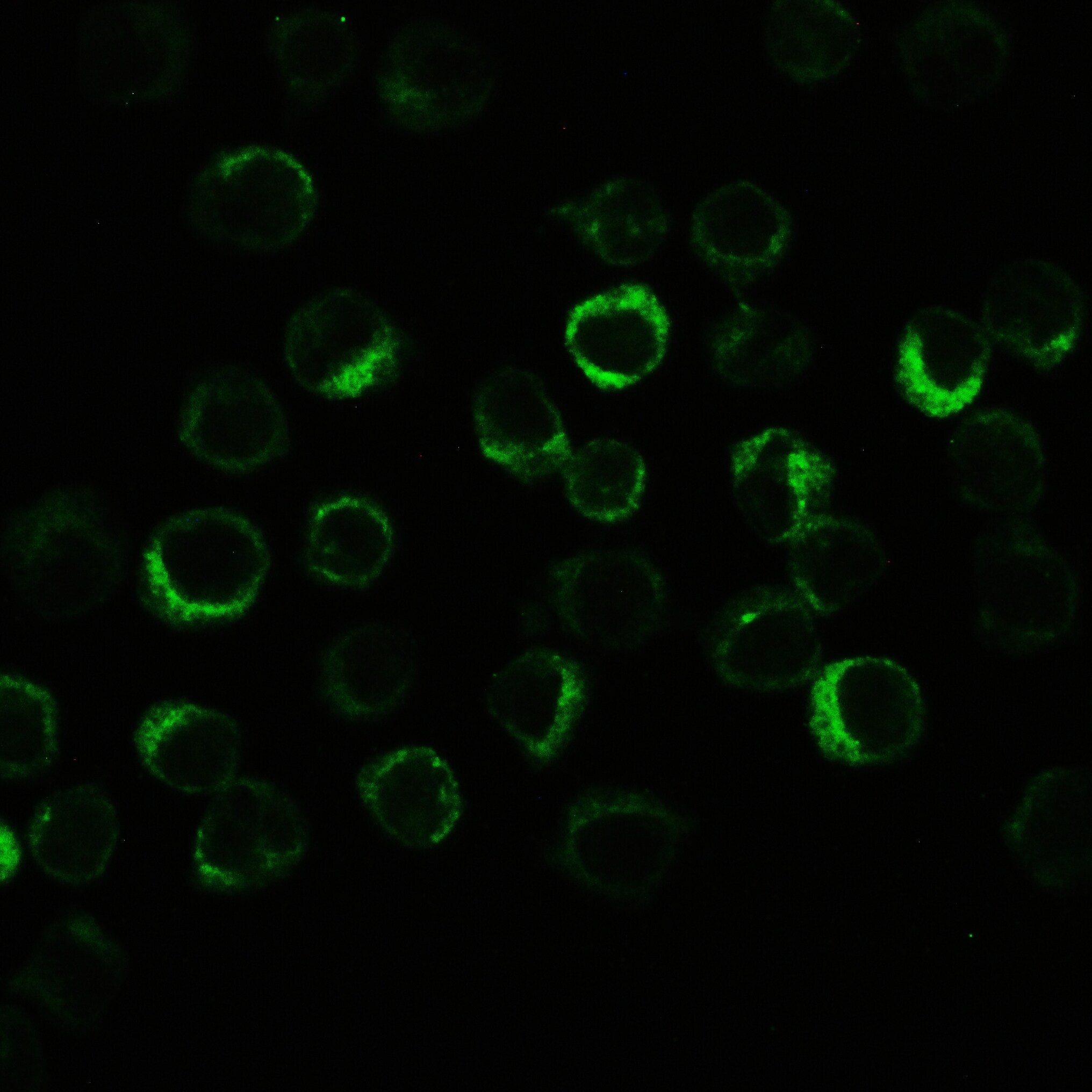
- mmunofluorescent analysis of NLRP3 on THP-1 cells. Cell Climbing Slice Preparation: Cover slip with cells was washed 3X in PBS and then immersed in 4% paraformaldehyde or neutral formalin for 10-20 minutes. Cover slip was washed 3X in PBS and allowed to dry for 8-10 hours. The cells were blocked with 10% goat serum and then incubated with 2 µg/mL NLRP3 polyclonal antibody (Product# PA5-79740) overnight at 4°C. DyLight ®488 Conjugated Goat Anti-Rabbit IgG was used as secondary antibody diluted 1:100 and incubated for 30 minutes at 37°C. Cells were visualized using a fluorescence microscope and filter sets appropriate for the label used.
- Submitted by
- Invitrogen Antibodies (provider)
- Main image
- Experimental details
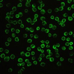
- mmunofluorescent analysis of NLRP3 on THP-1 cells. Cell Climbing Slice Preparation: Cover slip with cells was washed 3X in PBS and then immersed in 4% paraformaldehyde or neutral formalin for 10-20 minutes. Cover slip was washed 3X in PBS and allowed to dry for 8-10 hours. The cells were blocked with 10% goat serum and then incubated with 2 µg/mL NLRP3 polyclonal antibody (Product# PA5-79740) overnight at 4°C. DyLight ®488 Conjugated Goat Anti-Rabbit IgG was used as secondary antibody diluted 1:100 and incubated for 30 minutes at 37°C. Cells were visualized using a fluorescence microscope and filter sets appropriate for the label used.

- Submitted by
- Invitrogen Antibodies (provider)
- Main image
- Experimental details
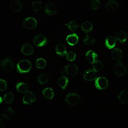
- mmunofluorescent analysis of NLRP3 on THP-1 cells. Cell Climbing Slice Preparation: Cover slip with cells was washed 3X in PBS and then immersed in 4% paraformaldehyde or neutral formalin for 10-20 minutes. Cover slip was washed 3X in PBS and allowed to dry for 8-10 hours. The cells were blocked with 10% goat serum and then incubated with 2 µg/mL NLRP3 polyclonal antibody (Product# PA5-79740) overnight at 4°C. DyLight ®488 Conjugated Goat Anti-Rabbit IgG was used as secondary antibody diluted 1:100 and incubated for 30 minutes at 37°C. Cells were visualized using a fluorescence microscope and filter sets appropriate for the label used.
Supportive validation
- Submitted by
- Invitrogen Antibodies (provider)
- Main image
- Experimental details
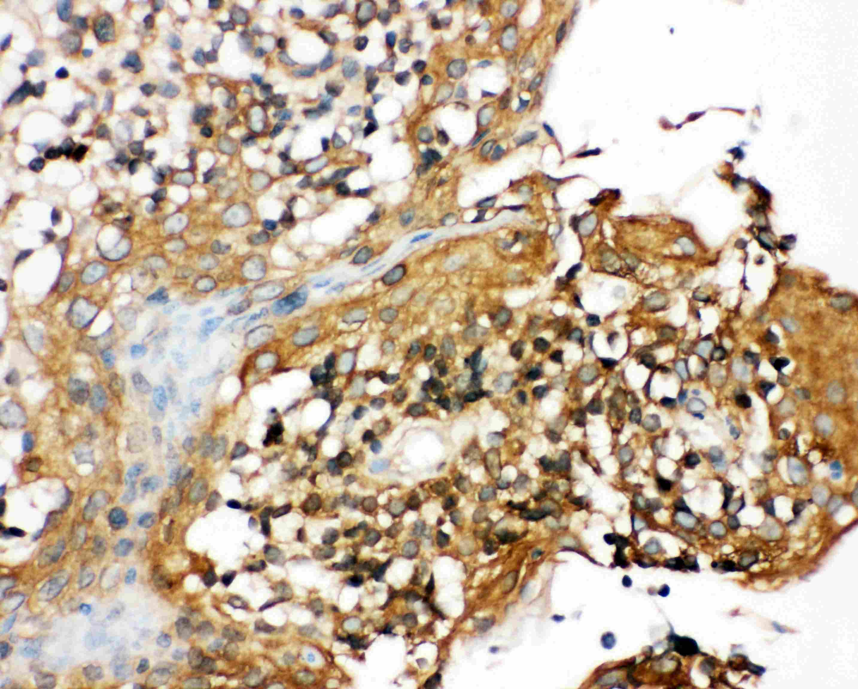
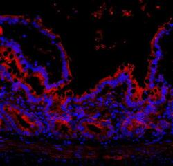
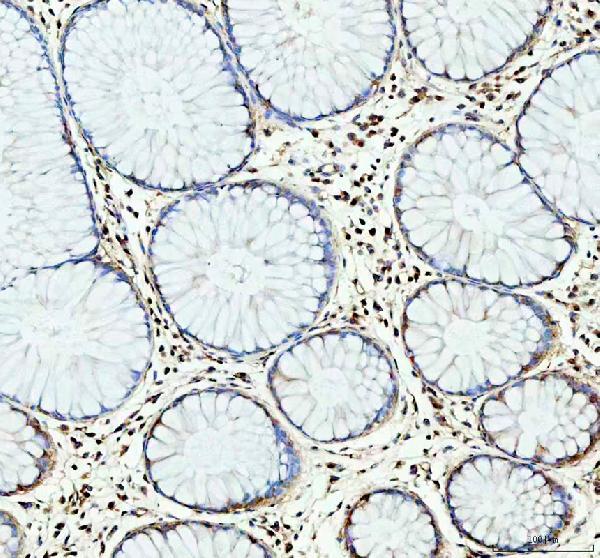
- Immunohistochemistry analysis of NLRP3 on paraffin-embedded human tonsil tissue. Antigen retrieval was performed using citrate buffer (pH6, epitope retrieval solution) for 20 mins. Sample was blocked using 10% goat serum, incubated with NLRP3 polyclonal antibody (Product# PA5-79740) with a dilution of 1 µg/mL (overnight at 4°C), and followed by biotinylated goat anti-rabbit IgG (30 minutes at 37°C). Development was performed using Streptavidin-Biotin-Complex (SABC) with DAB chromogen method.
- Submitted by
- Invitrogen Antibodies (provider)
- Main image

- Experimental details
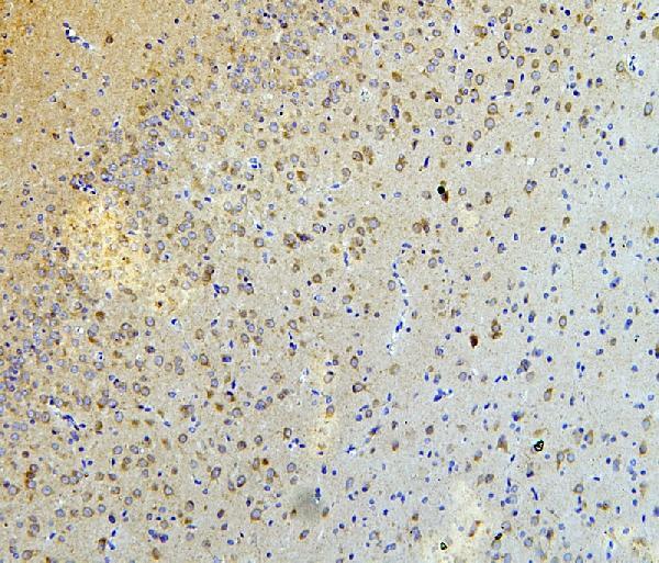
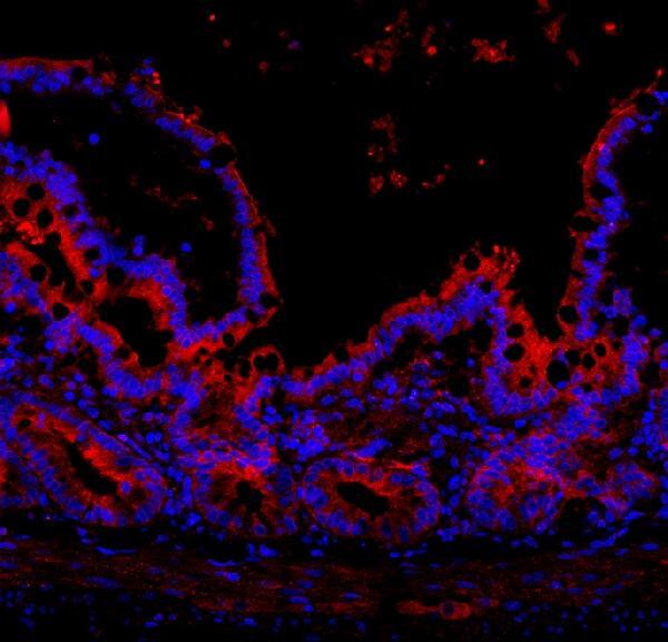
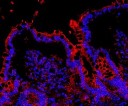
- Immunohistochemical analysis of NLRP3 in paraffin-embedded section of rat brain tissues. Heat mediated antigen retrieval was performed in citrate buffer (pH6, epitope retrieval solution) for 20 mins. The tissue section was blocked with 10% goat serum. The tissue section was then incubated with 1μg/mL rabbit anti-NLRP3 antibody (Product # PA5-79740) overnight at 4°C. Biotinylated goat anti-rabbit IgG was used as secondary antibody and incubated for 30 minutes at 37°C. The tissue section was developed using Strepavidin-Biotin-Complex (SABC) with DAB as the chromogen.
- Submitted by
- Invitrogen Antibodies (provider)
- Main image
- Experimental details
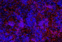
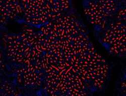
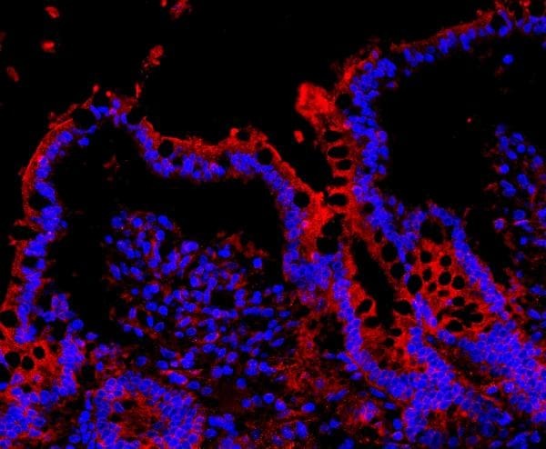
- Immunohistochemical analysis of CIAS1/NALP3 in paraffin-embedded section of rat colon tissues. Heat mediated antigen retrieval was performed in citrate buffer (pH6, epitope retrieval solution ) for 20 mins. The tissue section was blocked with 10% goat serum. The tissue section was then incubated with 1μg/mL rabbit anti-CIAS1/NALP3 antibody (Product # PA5-79740) overnight at 4°C. Cy3 Conjugated Goat Anti-Rabbit IgG was used as secondary antibody at 1:100 dilution and incubated for 30 minutes at 37°C. The section was counterstained with DAPI. Visualize using a fluorescence microscope and filter sets appropriate for the label used.
- Submitted by
- Invitrogen Antibodies (provider)
- Main image
- Experimental details
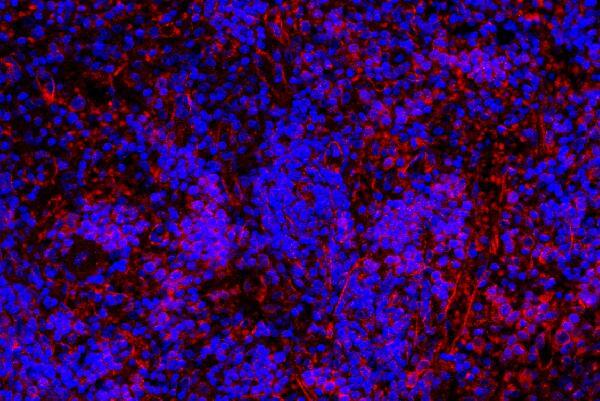
- Immunohistochemical analysis of CIAS1/NALP3 in paraffin-embedded section of rat spleen tissues. Heat mediated antigen retrieval was performed in citrate buffer (pH6, epitope retrieval solution ) for 20 mins. The tissue section was blocked with 10% goat serum. The tissue section was then incubated with 1μg/mL rabbit anti-CIAS1/NALP3 antibody (Product # PA5-79740) overnight at 4°C. Cy3 Conjugated Goat Anti-Rabbit IgG was used as secondary antibody at 1:100 dilution and incubated for 30 minutes at 37°C. The section was counterstained with DAPI. Visualize using a fluorescence microscope and filter sets appropriate for the label used.
- Submitted by
- Invitrogen Antibodies (provider)
- Main image
- Experimental details
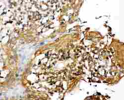
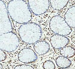
- Immunohistochemistry (Paraffin) analysis of NLRP3 in paraffin-embedded section of human colon cancer tissue using NLRP3 Polyclonal Antibody (Product # PA5-79740). Heat mediated antigen retrieval was performed in EDTA buffer (pH 8.0, epitope retrieval solution). The tissue section was blocked with 10% goat serum. The tissue section was then incubated with the primary antibody at a 2 µg/mL dilution overnight at 4°C. Peroxidase conjugated goat anti-rabbit IgG was used as secondary antibody and incubated for 30 minutes at 37°C. The tissue section was developed using HRP Conjugated Rabbit IgG Super Vision Assay Kit with DAB as the chromogen.
- Submitted by
- Invitrogen Antibodies (provider)
- Main image
- Experimental details
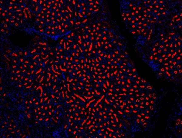
- Immunohistochemical analysis of CIAS1/NALP3 in paraffin-embedded section of rat kidney tissues. Heat mediated antigen retrieval was performed in citrate buffer (pH6, epitope retrieval solution ) for 20 mins. The tissue section was blocked with 10% goat serum. The tissue section was then incubated with 1μg/mL rabbit anti-CIAS1/NALP3 antibody (Product # PA5-79740) overnight at 4°C. Cy3 Conjugated Goat Anti-Rabbit IgG was used as secondary antibody at 1:100 dilution and incubated for 30 minutes at 37°C. The section was counterstained with DAPI. Visualize using a fluorescence microscope and filter sets appropriate for the label used.
- Submitted by
- Invitrogen Antibodies (provider)
- Main image
- Experimental details
- Immunohistochemical analysis of CIAS1/NALP3 in paraffin-embedded section of rat colon tissues. Heat mediated antigen retrieval was performed in citrate buffer (pH6, epitope retrieval solution ) for 20 mins. The tissue section was blocked with 10% goat serum. The tissue section was then incubated with 1μg/mL rabbit anti-CIAS1/NALP3 antibody (Product # PA5-79740) overnight at 4°C. Cy3 Conjugated Goat Anti-Rabbit IgG was used as secondary antibody at 1:100 dilution and incubated for 30 minutes at 37°C. The section was counterstained with DAPI. Visualize using a fluorescence microscope and filter sets appropriate for the label used.
Supportive validation
- Submitted by
- Invitrogen Antibodies (provider)
- Main image
- Experimental details
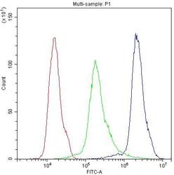
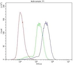
- Flow Cytometry of NLRP3 in THP-1 cells (blue line), isotype control rabbit IgG (green line) and unlabeled (red line). Samples were blocked with 10% goat serum, incubated with NLRP3 Polyclonal Antibody (Product # PA5-79740) at a dilution of 1 μg (per 1x10^6 cells), followed by DyLight®488 conjugated goat anti-rabbit IgG (for 30 minutes at 20°C) using 5-10 μg (per 1x10^6 cells) dilution.
- Submitted by
- Invitrogen Antibodies (provider)
- Main image

- Experimental details
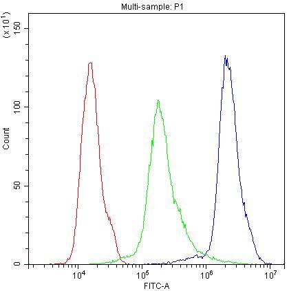
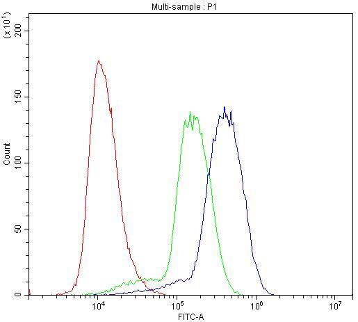
- Flow Cytometry of NLRP3 in U937 cells (blue line), isotype control rabbit IgG (green line) and unlabeled (red line). Samples were blocked with 10% goat serum, incubated with NLRP3 Polyclonal Antibody (Product # PA5-79740) at a dilution of 1 μg (per 1x10^6 cells), followed by DyLight®488 conjugated goat anti-rabbit IgG (for 30 minutes at 20°C) using 5-10 μg (per 1x10^6 cells) dilution.

- Submitted by
- Invitrogen Antibodies (provider)
- Main image

- Experimental details
- Flow Cytometry of NLRP3 in THP-1 cells (blue line), isotype control rabbit IgG (green line) and unlabeled (red line). Samples were blocked with 10% goat serum, incubated with NLRP3 Polyclonal Antibody (Product # PA5-79740) at a dilution of 1 μg (per 1x10^6 cells), followed by DyLight®488 conjugated goat anti-rabbit IgG (for 30 minutes at 20°C) using 5-10 μg (per 1x10^6 cells) dilution.

- Submitted by
- Invitrogen Antibodies (provider)
- Main image
- Experimental details
- Flow Cytometry of NLRP3 in U937 cells (blue line), isotype control rabbit IgG (green line) and unlabeled (red line). Samples were blocked with 10% goat serum, incubated with NLRP3 Polyclonal Antibody (Product # PA5-79740) at a dilution of 1 μg (per 1x10^6 cells), followed by DyLight®488 conjugated goat anti-rabbit IgG (for 30 minutes at 20°C) using 5-10 μg (per 1x10^6 cells) dilution.
- Submitted by
- Invitrogen Antibodies (provider)
- Main image

- Experimental details
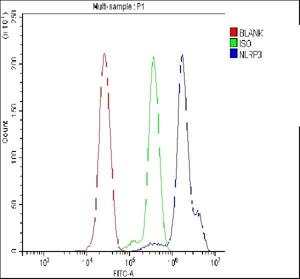
- Flow cytometry analysis of NLRP3 in THP-1 cells using NLRP3 Polyclonal Antibody (Product # PA5-79740), shown in overlay histogram (blue line). To facilitate intracellular staining, cells were fixed with 4% paraformaldehyde and permeabilized with permeabilization buffer. The cells were blocked with 10% normal goat serum, and incubated with the primary antibody (1 μg/1x10^6 cells) for 30 min at 20°C. DyLight 488 conjugated goat anti-rabbit IgG (5-10 µg/1x10^6 cells) was used as secondary antibody for 30 minutes at 20°C. Isotype control antibody (Green line) was rabbit IgG (1 µg/1x10^6) used under the same conditions. Unlabelled sample without incubation with primary antibody and secondary antibody (Red line) was used as a blank control.
Supportive validation
- Submitted by
- Invitrogen Antibodies (provider)
- Main image
- Experimental details
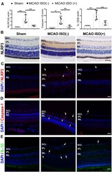
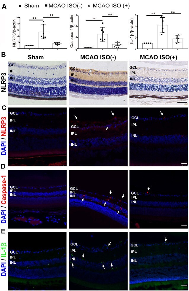
- Figure 4 ISO inhibits NLRP3 inflammasome activation in the retina after ischemic stroke in diabetic mice. (A) Relative gene expression of NLRP3, caspase-1, and IL-1beta in the retina. beta-Actin served as an endogenous reference gene ( n = 4 in Sham group; n = 6 in MCAO ISO (-) group and MCAO ISO (+) group). (B) Representative immunohistochemical staining for the protein expression of NLRP3 in the retina (magnification, 400x; scale bar, 50 mum). (C-E) Representative immunofluorescence staining for the protein expression of NLRP3 (C) , Caspase-1 (D) , and IL-1beta (E) in the retina. Arrow, NLRP3 (Red), Caspase-1 (Red), and IL-1beta (Green) positive cells (magnification, 400x; scale bar, 25 mum). GCL, ganglion cell layer; IPL, inner plexiform layer; INL, inner nuclear layer. * P < 0.05, ** P < 0.01.
- Submitted by
- Invitrogen Antibodies (provider)
- Main image
- Experimental details
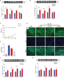
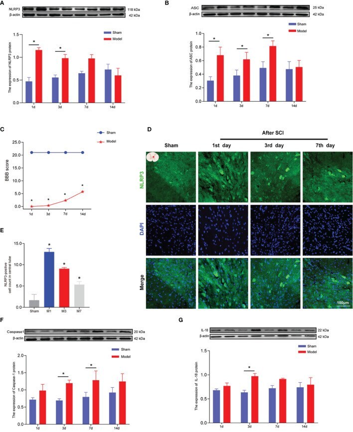
- The expression levels of NLRP3, ASC, caspase-1, and IL-18 in rat spinal cords 1, 3, 7, and 14 days after SCI. (A) Representative Western blotting and semiquantitative analysis of NLRP3 protein in the spinal cords of rats in the sham and model groups at days 1, 3, 7, and 14 post-SCI. The image shows representative Western blotting for NLRP3 and beta-actin. (B) Western blotting was used to analyze the protein expression of ASC in the sham and model groups after SCI. Representative protein bands are shown for ASC at 1, 3, 7, and 14 days after SCI (N = 5 rats/group). The expression of ASC is shown for the model and sham groups. *p < 0.05; two-way ANOVA. (C) The BBB function scores in the sham and model groups. N = 8 rats/group. *p < 0.05. (D) The expression of NLRP3 in the spinal cords of rats was detected using IF. The expression of NLRP3 in the spinal cord was detected in the sham group and 1, 3, and 7 days after SCI. Cells positive for NLRP3 protein are shown in green. (E) Quantitative analysis of NLRP3-positive cells in the model group compared with the sham group. *p < 0.05; one-way ANOVA was used for comparison. N = 5 rats/group. (F, G) Protein expression and representative protein bands for caspase-1 and IL-18 at each time point after SCI as determined using Western blotting. N = 5 rats/group. *p < 0.05; two-way ANOVA was used for comparison. Data are presented as the mean +- SEM. SCI, spinal cord injury; BBB, Basso-Beattie-Bresnahan; IF, immunofluorescence.
- Submitted by
- Invitrogen Antibodies (provider)
- Main image
- Experimental details
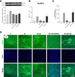
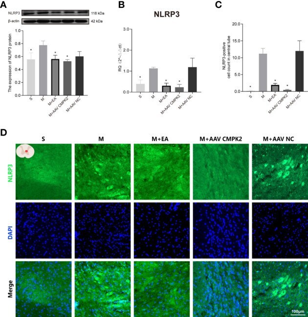
- Effects of CMPK2 knockdown on activation of the NLRP3 inflammasome in rat spinal cord. (A) Western blotting analysis of NLRP3 expression in spinal cords of rats in the Sham group, Model, M+EA, M+AAV CMPK2, and M+AAV NC groups. A representative Western blotting and semiquantitative analysis of NLRP3 protein levels are shown. N = 5 rats/group. *p < 0.05 compared with the Model group; one-way anOVA was used for comparison. (B) Quantitative PCR analysis of NLRP3 gene expression in the spinal cords of rats in the Sham, Model, M+EA, M+AAV CMPK2, and M+AAV NC groups. N = 5 rats in each group. *p < 0.05 compared with the model group; one-way anOVA was used for comparison. (C) Quantitative analysis of NLRP3-positive cells. *p < 0.05 compared with the model group; one-way anOVA was used for comparison. N = 5 rats/group. (D) The expression of NLRP3 in the spinal cords of rats in the Sham, Model, M+EA, M+AAV CMPK2, and M+AAV NC groups were detected using IF. The number of CMPK2-positive cells (green fluorescence) 3 days after SCI was labeled by immunofluorescence, and the areas with positive NLRP3 staining are shown in green. All data are expressed as the mean +- SEM. IF, immunofluorescence; SCI, spinal cord injury.
- Submitted by
- Invitrogen Antibodies (provider)
- Main image
- Experimental details
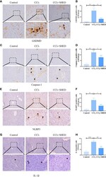
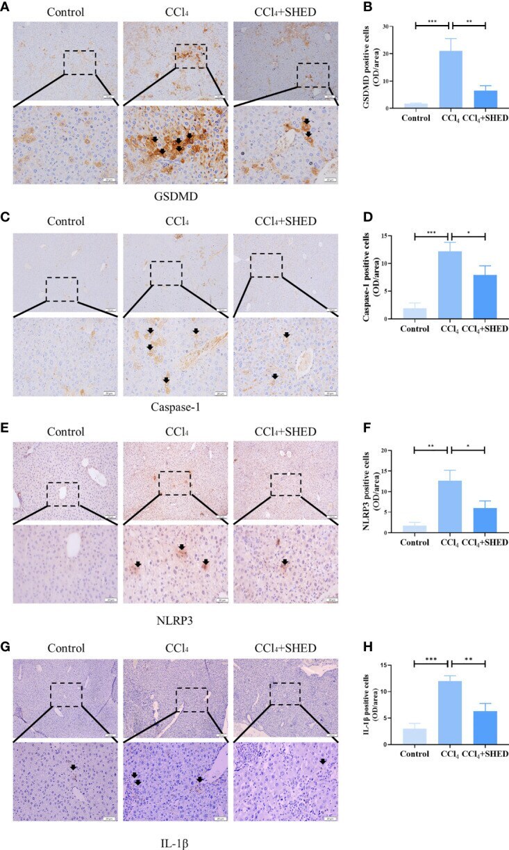
- Figure 2 SHED attenuated pyroptosis of hepatocytes in CCl 4 -induced liver cirrhosis. (A, B) Immunohistochemical staining of GSDMD in control, CCl 4 , and SHED transplantation groups (C-H) . The expression of NLRP3, Caspase-1, and IL-1beta in control, CCl 4 , and SHED transplantation groups, as assessed by immunohistochemical staining. Scale bar: 100 and 20 mum. *p < 0.05, **p < 0.01, and ***p < 0.001. Graph bars show the mean +- SD. All of the assays were performed in triplicate. NLRP3, NOD-like receptor family pyrin domain containing 3; GSDMD, gasdermin D; SHED, stem cells from human exfoliated deciduous teeth.
- Submitted by
- Invitrogen Antibodies (provider)
- Main image

- Experimental details
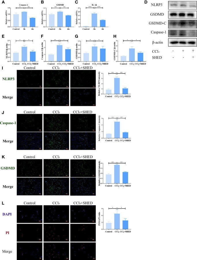
- Figure 3 SHED inhibited CCl 4 -induced NLRP3 inflammasome activation and pyroptosis in hepatocytes. (A-C) The mRNA level of Caspase-1, GSDMD, and IL-1b was detected after 3h and 6h CCl 4 stimulation analyzed by RT-qPCR. (D-H) The expression levels of NLRP3, Caspase-1, and GSDMD-C in hepatocytes in control, CCl 4 , and SHED infusion group by Western blotting analysis. (I-K) THe expression of NLRP3, Caspase-1 and GSDMD in hepatocytes in control, CCl 4 , and CCl 4 +SHED groups was analyzed by immunofluorescence staining. (L) The ratio of dead cells was determined by PI staining in control, CCl 4 , and CCl 4 +SHED groups. Scale bar: 50 mum. *p < 0.05, **p < 0.01, and ***p < 0.001. Graph bars show the mean +- SD. All of the assays were performed in triplicate. PI, propidium iodide; SHED, stem cells from human exfoliated deciduous teeth; GSDMD, gasdermin D.
- Submitted by
- Invitrogen Antibodies (provider)
- Main image
- Experimental details
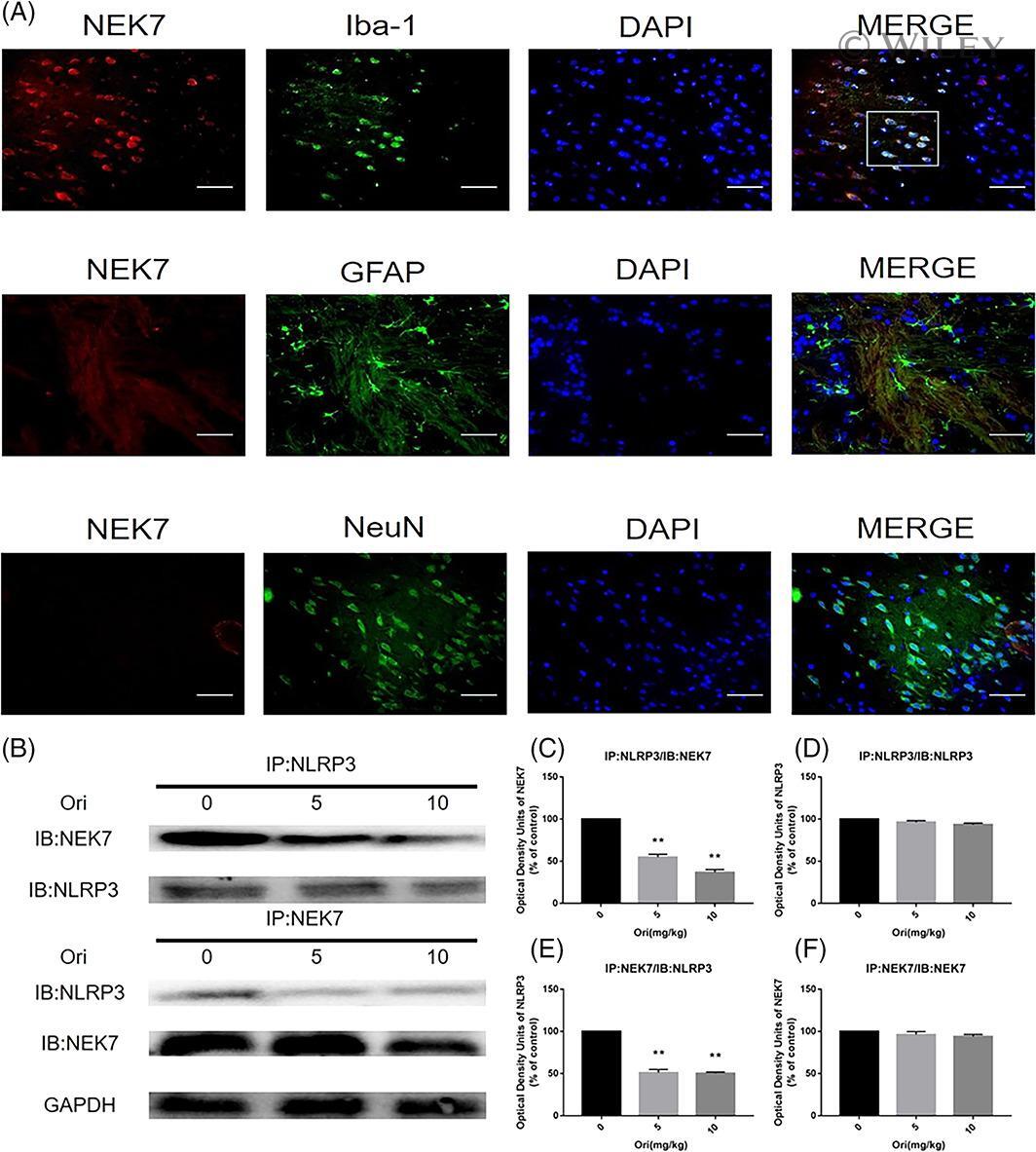
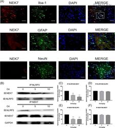
- 7 FIGURE Detection of the interaction between NLRP3 and NEK7 in hippocampus. (a) Immunofluorescent double staining. (b) Co-immunoprecipitation of NEK7 and NLRP3. (c-f) Western blot analysis of NEK7 and NLRP3. The data (mean +- SEM) were analyzed by Student's t -test for two-group comparisons, n = 3-4. The experiment was repeated three times. ** p < .001 versus Control group