 Explore
 Validate
 Learn
Learn Western blot
 Flow cytometry
Antibody data
- Antibody Data
- Antigen structure
- References [4]
- Comments [0]
- Validations
- Western blot [2]
- Immunocytochemistry [1]
- Immunohistochemistry [1]
- Other assay [1]
Submit
Validation data
Reference
Comment
Report error
- Product number
- 14-9773-80 - Provider product page

- Provider
- Invitrogen Antibodies
- Product name
- Anti-SOX11 Monoclonal Antibody (SOX11-C1), eBioscience™
- Antibody type
- Monoclonal
- Antigen
- Other
- Description
- Description: The monoclonal antibody SOX11-C1 (C1) recognizes human Sox11. The transcription factor Sox11 is a member of the SOX (sex determining region Y-related HMG (High Mobility Group) Box) family of proteins. Under normal conditions Sox11 is expressed in the developing central nervous system during embryogenesis and has sequence homology to Sox4. Sox11 plays a role in neuronal maturation and epithelial-mesenchymal interactions and is required for the survival of neuronal and mesenchymal progenitor cells. Sox11 is expressed in mantle cell lymphoma (MCL) and in subsets of hairy cell leukemias, Burkitt lymphomas, and B cell lymphoblastic leukemias, but is not expressed in other B cell lymphomas or in normal B lymphocytes. Sox11 is also expressed in epithelial ovarian cancer and gliomas. Applications Reported: This SOX11-C1 antibody has been reported for use in flow cytometric analysis, western blotting, immunohistochemical staining of formalin-fixed paraffin embedded tissue sections, and immunocytochemistry. Applications Tested: This SOX11-C1 antibody has been tested by immunohistochemistry of formalin-fixed paraffin embedded tissue using low or high pH antigen retrieval. The antibody can be used at less than or equal to 5 µg/mL. This SOX11-C1 antibody has been tested by immunocytochemistry of fixed and permeabilized cells and can be used at less than or equal to 5 µg/mL. It is recommended that the antibody be carefully titrated for optimal performance in the assay of interest. Purity: Greater than 90%, as determined by SDS-PAGE. Aggregation: Less than 10%, as determined by HPLC. Filtration: 0.2 µm post-manufacturing filtered.
- Reactivity
- Human
- Host
- Mouse
- Isotype
- IgG
- Antibody clone number
- SOX11-C1
- Vial size
- 25 µg
- Concentration
- 0.5 mg/mL
- Storage
- 4° C
Submitted references The role of SOX11 in mantle cell lymphoma.
Expanded clinical and experimental use of SOX11 - using a monoclonal antibody.
Nuclear expression of the non B-cell lineage Sox11 transcription factor identifies mantle cell lymphoma.
Expression of the Sox11 gene in mouse embryos suggests roles in neuronal maturation and epithelio-mesenchymal induction.
Lu TX, Li JY, Xu W
Leukemia research 2013 Nov;37(11):1412-9
Leukemia research 2013 Nov;37(11):1412-9
Expanded clinical and experimental use of SOX11 - using a monoclonal antibody.
Nordström L, Andréasson U, Jerkeman M, Dictor M, Borrebaeck C, Ek S
BMC cancer 2012 Jun 27;12:269
BMC cancer 2012 Jun 27;12:269
Nuclear expression of the non B-cell lineage Sox11 transcription factor identifies mantle cell lymphoma.
Ek S, Dictor M, Jerkeman M, Jirström K, Borrebaeck CA
Blood 2008 Jan 15;111(2):800-5
Blood 2008 Jan 15;111(2):800-5
Expression of the Sox11 gene in mouse embryos suggests roles in neuronal maturation and epithelio-mesenchymal induction.
Hargrave M, Wright E, Kun J, Emery J, Cooper L, Koopman P
Developmental dynamics : an official publication of the American Association of Anatomists 1997 Oct;210(2):79-86
Developmental dynamics : an official publication of the American Association of Anatomists 1997 Oct;210(2):79-86
No comments: Submit comment
Supportive validation
- Submitted by
- Invitrogen Antibodies (provider)
- Main image

- Experimental details
- Knockdown of SOX11 was achieved by transfecting SH-SY5Y with SOX11 specific siRNAs (Silencer® select Product # s224668, s194808) . Western blot analysis (Fig. a) was performed using modified whole cell extracts (1% SDS) from the SOX11 knockdown cells (Lane 3), non-specific scrambled siRNA transfected cells (Lane 2) and untransfected cells (Lane 1). The blot was probed with SOX11 Monoclonal Antibody (SOX11-C1), eBioscience™ (Product # 14-9773-82, 1:1000 dilution) and Goat anti-Mouse IgG (H+L) Superclonal™ Recombinant Secondary Antibody, HRP (Product # A28177, 1:4000 dilution). Densitometric analysis of this western blot is shown i histogram (Fig. b). Loss of signal upon siRNA mediated knock down confirms that antibody is specific to SOX11.

- Submitted by
- Invitrogen Antibodies (provider)
- Main image

- Experimental details
- Western blot was performed using SOX11 Monoclonal Antibody (SOX11-C1), eBioscience™ (Product # 14-9773-82) and a 55 kDa band corresponding to SOX11 was observed across cell lines tested except BeWo and HL-60 which are reported to be negative. Whole cell extracts (30 µg lysate) of SHSY-5Y (Lane 1), IMR-32 (Lane 2), BeWo (Lane 3) HL-60 (Lane 4) were electrophoresed using NuPAGE® 4-12 % Bis-Tris gel (Product # NP0321BOX). Resolved proteins were then transferred onto a nitrocellulose membrane (Product # IB23001) by iBlot® 2 Dry Blotting System (Product # IB21001). The blot was probed with the primary antibody (1:1000 dilution) and detected by chemiluminescence with Goat anti-Mouse IgG (H+L) Superclonal™ Recombinant Secondary Antibody, HRP (Product # A28177, 1:4000 dilution) using the iBright FL 1000 (Product # A32752). Chemiluminescent detection was performed using Novex® ECL Chemiluminescent Substrate Reagent Kit (Product # WP20005)..

Supportive validation
- Submitted by
- Invitrogen Antibodies (provider)
- Main image

- Experimental details
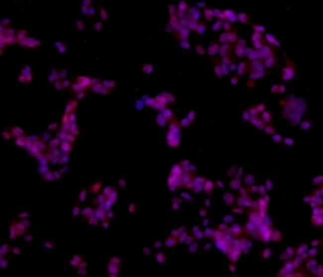
- Immunocytochemistry of fixed and permeabilized HepG2 cells using 10 µg/mL of Anti-Human Sox11 Purified followed by F (ab')2 Anti-Mouse IgG eFluor® 570.Nuclei are stained with DAPI, colocalization appears pink.
Supportive validation
- Submitted by
- Invitrogen Antibodies (provider)
- Main image

- Experimental details
- Immunohistochemistry of formalin-fixed paraffin embedded human Mantle Cell lymphoma using 10 µg/mL of Anti-Human Sox11 Purified followed by Anti-Mouse IgG Biotin, Streptavidin HRP, and DAB visualization.Nuclei are counterstained with hematoxylin.

Supportive validation
- Submitted by
- Invitrogen Antibodies (provider)
- Main image

- Experimental details
- Immunofluorescence analysis of SOX11 was performed using 70% confluent log phase SHSY-5Y and BeWo cells. The cells were fixed with 4% paraformaldehyde for 10 minutes, permeabilized with 0.1% Triton™ X-100 for 15 minutes, and blocked with 2% BSA for 1 hour at room temperature. The cells were labeled with SOX11 Monoclonal Antibody (SOX11-C1), eBioscience™ (Product # 14-9773-82) at 5µg/ml concentration in 0.1% BSA, incubated at 4 degree Celsius overnight and then labeled with Goat anti-Rabbit IgG (H+L) Superclonal™ Recombinant Secondary Antibody, Alexa Fluor® 488 (Product # A27034) at a dilution of 1:2000 for 45 minutes at room temperature (Panel a: green). Nuclei (Panel b: blue) were stained with SlowFade® Gold Antifade Mountant with DAPI (Product # S36938). F-actin (Panel c: red) was stained with Rhodamine Phalloidin (Product # R415, 1:300). Panel d represents the merged image showing localization to nucleus. Panel e shows BeWo cells with no expression of SOX11. Panel f represents control cells with no primary antibody to assess background. The images were captured at 60X magnification.
