Explore
 Validate
 Learn
Learn Immunohistochemistry
 Flow cytometry
Antibody data
- Antibody Data
- Antigen structure
- References [1]
- Comments [0]
- Validations
- Immunohistochemistry [2]
- Other assay [4]
Submit
Validation data
Reference
Comment
Report error
- Product number
- 50-9773-80 - Provider product page

- Provider
- Invitrogen Antibodies
- Product name
- SOX11 Monoclonal Antibody (SOX11-C1), eFluor™ 660, eBioscience™
- Antibody type
- Monoclonal
- Antigen
- Other
- Description
- Description: The monoclonal antibody SOX11-C1 (C1) recognizes human Sox11. The transcription factor Sox11 is a member of the SOX (sex determining region Y-related HMG (High Mobility Group) Box) family of proteins. Under normal conditions Sox11 is expressed in the developing central nervous system during embryogenesis and has sequence homology to Sox4. Sox11 plays a role in neuronal maturation and epithelial-mesenchymal interactions and is required for the survival of neuronal and mesenchymal progenitor cells. Sox11 is expressed in mantle cell lymphoma (MCL) and in subsets of hairy cell leukemias, Burkitt lymphomas, and B cell lymphoblastic leukemias, but is not expressed in other B cell lymphomas or in normal B lymphocytes. Sox11 is also expressed in epithelial ovarian cancer and gliomas. Applications Reported: This SOX11-C1 antibody has been reported for use in immunohistochemical staining of formalin-fixed paraffin embedded tissue sections, immunocytochemistry and flow cytometric analysis. Applications Tested: This SOX11-C1 antibody has been tested by immunohistochemistry of formalin-fixed paraffin embedded human tissue using low or high pH antigen retrieval and can be used at less than or equal to 10 µg/mL. It is recommended that the antibody be carefully titrated for optimal performance in the assay of interst. eFluor® 660 is a replacement for Alexa Fluor® 647. eFluor® 660 emits at 659 nm and is excited with the red laser (633 nm). Please make sure that your instrument is capable of detecting this fluorochome. Excitation: 633-647 nm; Emission: 668 nm; Laser: Red Laser. Filtration: 0.2 µm post-manufacturing filtered.
- Reactivity
- Human
- Host
- Mouse
- Isotype
- IgG
- Antibody clone number
- SOX11-C1
- Vial size
- 25 µg
- Concentration
- 0.2 mg/mL
- Storage
- 4° C, store in dark, DO NOT FREEZE!
Submitted references SOX11 promotes epithelial/mesenchymal hybrid state and alters tropism of invasive breast cancer cells.
Oliemuller E, Newman R, Tsang SM, Foo S, Muirhead G, Noor F, Haider S, Aurrekoetxea-Rodríguez I, Vivanco MD, Howard BA
eLife 2020 Sep 10;9
eLife 2020 Sep 10;9
No comments: Submit comment
Supportive validation
- Submitted by
- Invitrogen Antibodies (provider)
- Main image

- Experimental details
- Immunohistochemistry of formalin-fixed paraffin embedded human Mantle Cell lymphoma using 10 µg/mL of Mouse IgG1 K Isotype Control eFluor® 660 (left) or 10 µg/mL of Anti-Human Sox11 eFluor® 660 (right). Nuclei are stained with DAPI (colocalization appears pink).

Supportive validation
- Submitted by
- Invitrogen Antibodies (provider)
- Main image
- Experimental details
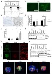
- Figure 1. Inducible expression of SOX11 leads to changes in cell state profiles of DCIS.com cells. ( A ) Western blot of SOX11 in cytoplasmic and nuclear fractions of DCIS.com cells containing the pInducer21 empty vector in presence (iEV) or absence (niEV) of 1 muM Doxycycline (DOX) or the pInducer21SOX11 with (iSOX11) or without DOX (niSOX11). GAPDH and LAMIN B1 were used as loading control of cytoplasmic and nuclear fractions, respectively. Densitometry results normalised against niSOX11 are shown in brackets. ( B ) SOX11 expression detected in iSOX11 cells stained by IF after 48 hr of DOX induction. Scale Bar: 200 mum. ( C ) ER- DCIS case sample showing SOX11 staining in DCIS and adjacent normal breast tissue. Scale Bar: 200 mum. ( D ) Results from flow cytometry analysis of Aldefluor assays of niEV and niSOX11 cells (day 0) and iEV and iSOX11 after 2 days treatment with 1 muM DOX. Results show the % of ALDH+ cells normalised against niEV. Error bars represent SD. *p = 0.0223. n = 5. ( E ) Results from flow cytometry analysis of CD24 and CD44 of niEV and niSOX11 cells (day 0) and iEV and iSOX11 after treatment with 1 muM DOX for 2 days. Results show the average % of cells CD44+/CD24+ in each condition. Error bars represent SD. ***p = 0.0005 (iSOX11 vs niSOX11) and p = 0.0009 (iSOX11 vs iEV) n = 3. ( F ) Western blot of CD24 in cytoplasmic and nuclear fractions of niEV, niSOX11, iEV and iSOX11 cells. GAPDH and LAMIN B1 were used as loading control of cytoplasmic and nuclear
- Submitted by
- Invitrogen Antibodies (provider)
- Main image

- Experimental details
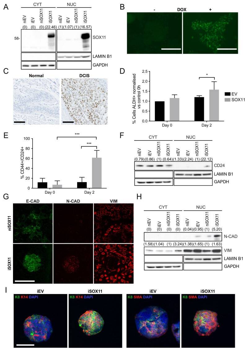
- Figure 3. SOX11 expression promotes expression of developmental pathways frequently activated in cancer. ( A ) Volcano plot representing the RNAs with a log2 fold-change > +/- 0.585 in the RNA-sequencing results of iSOX11 cells grown in 2D compared with the controls [(iSOX11-niSOX11)- (iEV-niEV)] to account for effects of DOX treatment on DCIS.com cells. ( B ) Gene ontology results from A. ( C ) List of genes overexpressed log2 fold-change >+/-1.585 times in all three RNA-sequencing (cells grown in: 2D, 3D for 2 days, 3D for 5 days) results comparing iSOX11 versus iEV. ( D ) qRT-PCR results for several potential SOX11 targets in EV and SOX11 cells with and without DOX induction in cells grown in 2D. Experiment was repeated three times. ( E ) Western blot of MEX3A and TUBB3 in cytoplasmic and nuclear fractions of EV or SOX11 cells in presence or absence of 1 muM DOX. GAPDH and LAMIN B1 were used as loading control of cytoplasmic and nuclear fractions respectively. In brackets, densitometry results normalised against niEV and niSOX11. ( F ) IF staining of DCIS iEV and DCIS iSOX11 cells with TUBB3 (green) and MEX3A (red). Scale: 200 mum. ( G ) Western blot of MEX3A and TUBB3 in SOX11+ breast cancer cell lines and SOX11- DCIS.com and MCF10A from the MCF10A mammary cell progression series. ( H ) Pie charts representing the percentage of breast cancer samples with a log2 fold-change greater than two in the levels of MEX3A or TUBB3 RNA when SOX11 increased between 0.5- and 2-fold, 2
- Submitted by
- Invitrogen Antibodies (provider)
- Main image
- Experimental details
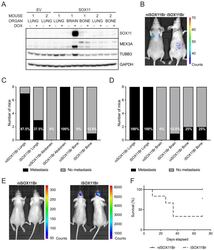
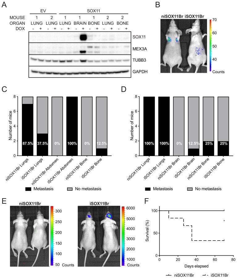
- Figure 5. SOX11+ DCIS cells isolated from brain metastasis display a colonisation and growth advantage after intracranial xenografting. ( A ) Western blot of SOX11, MEX3A and TUBB3 in total cell lysates of EV and SOX11 cell lines isolated from primary metastasis at indicated sites in presence or absence of DOX. ( B ) Representative in vivo IVIS imaging 7 days after tail vein injections of iSOX11 cells that were isolated from the brain metastasis (SOX11Br) in presence or absence of DOX. ( C ) Tabulated results of micrometastasis from in vivo IVIS imaging 7 days after tail vein injections of SOX11Br cells. ( D ) Tabulated results of micrometastasis from ex vivo IVIS imaging of the tail vein injections of SOX11Br cells. ( E ) IVIS imaging of mice fed normal chow or DOX-containing chow 10 days after intracranial injections of SOX11Br cells. ( F ) Survival curve for mice shown in E. *p = 0.0195. DOX: doxycycline.
- Submitted by
- Invitrogen Antibodies (provider)
- Main image

- Experimental details
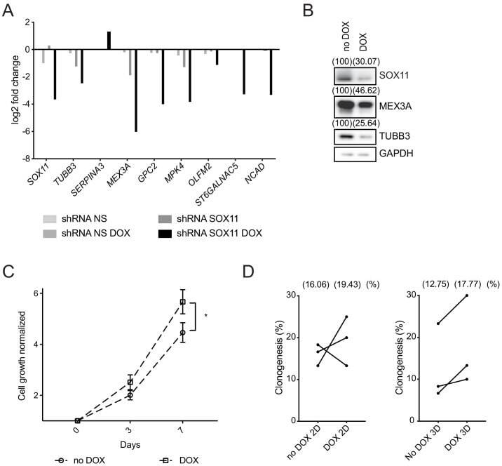
- Figure 6. SOX11 regulates growth of ER- breast cancer cells. ( A ) qRT-PCR results for several potential SOX11 targets in CAL-148 cells transduced with shRNA to SOX11 or shRNA NS cells with and without DOX induction in cells grown in 2D. ( B ) Western blot of SOX11, MEX3A and TUBB3 in total cell lysates of CAL-148 cells transduced with shRNA SOX11 in presence or absence of 1 muM DOX after 48 hr. GAPDH was used as loading control. Densitometry results normalised against no DOX are shown in brackets. ( C ) Cell growth assay results for CAL-148 shRNA SOX11 cells induced with 1 muM DOX at 3 and 7 days. Experiments were performed three times. Error bars represent SEM. *p=0.0106 (day 7). ( D ) Quantification of clonogenicity in 2D and 3D from single CAL-148 shRNA SOX11 cells plated in presence or absence of DOX after 21 days. The number in brackets represents the mean in each group of the three experimental replicates. DOX: doxycycline, NS: non-silencing.