 Explore
 Validate
 Learn
Learn Western blot
Antibody data
- Antibody Data
- Antigen structure
- References [0]
- Comments [0]
- Validations
- Western blot [1]
- Immunocytochemistry [3]
Submit
Validation data
Reference
Comment
Report error
- Product number
- PA5-29482 - Provider product page

- Provider
- Invitrogen Antibodies
- Product name
- SRC2 Polyclonal Antibody
- Antibody type
- Polyclonal
- Antigen
- Recombinant protein fragment
- Description
- Recommended positive controls: HeLa Nucleus. Predicted reactivity: Mouse (89%), Rat (93%), Pig (95%). Store product as a concentrated solution. Centrifuge briefly prior to opening the vial.
- Reactivity
- Human
- Host
- Rabbit
- Isotype
- IgG
- Vial size
- 100 µL
- Concentration
- 1 mg/mL
- Storage
- Store at 4°C short term. For long term storage, store at -20°C, avoiding freeze/thaw cycles.
No comments: Submit comment
Supportive validation
- Submitted by
- Invitrogen Antibodies (provider)
- Main image

- Experimental details
- Western Blot using SRC2 Polyclonal Antibody (Product # PA5-29482). Sample (20 µg). Lane A: HeLa Nucleus. 5% SDS PAGE. SRC2 Polyclonal Antibody (Product # PA5-29482) diluted at 1:500. The HRP-conjugated anti-rabbit IgG antibody was used to detect the primary antibody.

Supportive validation
- Submitted by
- Invitrogen Antibodies (provider)
- Main image

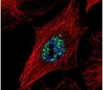
- Experimental details
- Immunofluorescent analysis of NCOA2 in paraformaldehyde-fixed HeLa cells using a NCOA2 polyclonal antibody (Product # PA5-29482) (Green) at a 1:500 dilution. Alpha-tubulin filaments were labeled with Product # PA5-29281 (Red) at a 1:2000.
- Submitted by
- Invitrogen Antibodies (provider)
- Main image
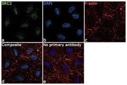
- Experimental details
- Immunofluorescence analysis of SRC2 was performed using 70% confluent log phase A549 cells. The cells were fixed with 4% paraformaldehyde for 10 minutes, permeabilized with 0.1% Triton™ X-100 for 15 minutes, and blocked with 1% BSA for 1 hour at room temperature. The cells were labeled with SRC2 Polyclonal Antibody (Product # PA5-29482) at 5 µg/mL concentration in 0.1% BSA, incubated at 4 degree Celsius overnight and then labeled with Goat anti-Rabbit IgG (H+L) Superclonal™ Secondary Antibody, Alexa Fluor® 488 conjugate (Product # A27034) for 45 minutes at room temperature (Panel a: green). Nuclei (Panel b: blue) were stained with ProLong™ Diamond Antifade Mountant with DAPI (Product # P36962). F-actin (Panel c: red) was stained with Rhodamine Phalloidin (Product # R415, 1:300). Panel d represents the merged image showing nuclear localization. Panel e represents control cells with no primary antibody to assess background. The images were captured at 60X magnification.

- Submitted by
- Invitrogen Antibodies (provider)
- Main image

- Experimental details
- Knockdown of SCR2 was achieved by transfecting A549 cells with SRC2 specific siRNA (Silencer® select Product # s20581, s20582). Immunofluorescence analysis was performed on A549 cells (untransfected, panel a,d), transfected with non-specific scrambled siRNA (panels b,e) and transfected with SRC2 specific siRNA (panel c,f) Cells were fixed, permeabilized, and labelled with SRC2 Polyclonal Antibody (Product # PA5-29482, 5 µg/mL), followed by Goat anti-Rabbit IgG (H+L) Superclonal™ Secondary Antibody, Alexa Fluor® 488 conjugate (Product # A27034, 1:2000). Nuclei (blue) were stained using ProLong™ Diamond Antifade Mountant with DAPI (Product # P36962), and Rhodamine Phalloidin (Product # R415, 1:300) was used for cytoskeletal F-actin (red) staining. Reduction of specific signal was observed upon siRNA mediated knockdown (panel c,f) confirming specificity of the antibody to SRC2 (green). The images were captured at 60X magnification
