Explore
 Validate
 Learn
LearnMA1-19385
antibody from Invitrogen Antibodies
Targeting: CLU
APOJ, CLI, CLU1, CLU2, KUB1, SGP-2, SP-40, TRPM-2
 Western blot
 ELISA
ELISAAntibody data
- Antibody Data
- Antigen structure
- References [0]
- Comments [0]
- Validations
- Western blot [3]
- Immunocytochemistry [1]
Submit
Validation data
Reference
Comment
Report error
- Product number
- MA1-19385 - Provider product page

- Provider
- Invitrogen Antibodies
- Product name
- Apolipoprotein J Monoclonal Antibody (Hs-3)
- Antibody type
- Monoclonal
- Antigen
- Other
- Description
- This antibody will not cross-react with porcine, bovine, canine or feline.
- Reactivity
- Human
- Host
- Mouse
- Isotype
- IgG
- Antibody clone number
- Hs-3
- Vial size
- 100 µg
- Concentration
- 1 mg/mL
- Storage
- 4° C, do not freeze
No comments: Submit comment
Supportive validation
- Submitted by
- Invitrogen Antibodies (provider)
- Main image
- Experimental details
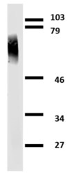
- Western blotting analysis of human clusterin using mouse monoclonal antibody Hs-3 on lysates of MCF-7 cell line and MOLT-4 cell line (clusterin non-expressing cell line; negative control) under non-reducing and reducing conditions. Nitrocellulose membrane was probed with 2µg/mL of mouse anti-clusterin monoclonal antibody (Product # MA1-19385) followed by IRDye800-conjugated anti-mouse secondary antibody. Clusterin was detected around 40kDa.

- Submitted by
- Invitrogen Antibodies (provider)
- Main image

- Experimental details
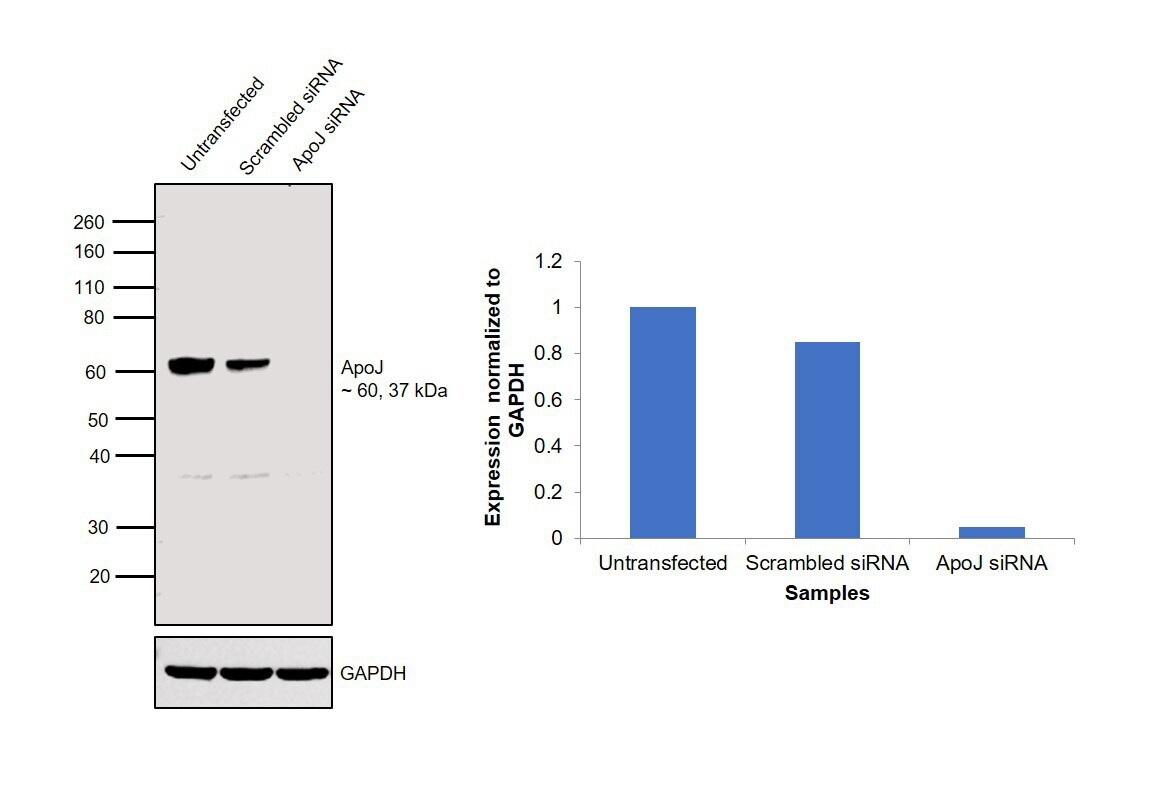
- Knockdown of Apolipoprotein J was achieved by transfecting HeLa cells with Apolipoprotein J specific siRNAs (Silencer® select Product # s3156, s3158). Western blot analysis (Fig. a) was performed using whole cell extracts from the HeLa knockdown cells (lane 3), non-specific scrambled siRNA transfected cells (lane 2) and untransfected cells (lane 1). The blot was probed with Apolipoprotein J Mouse Monoclonal Antibody (Product # MA1-19385, 1 µg/mL) and Goat anti-Mouse IgG (H+L) Superclonal™ Recombinant Secondary Antibody, HRP (Product # A28177, 1:4000 dilution). Densitometric analysis of this western blot is shown in histogram (Fig. b). Decrease in signal upon siRNA mediated knock down confirms that antibody is specific to Apolipoprotein J.
- Submitted by
- Invitrogen Antibodies (provider)
- Main image
- Experimental details
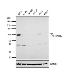
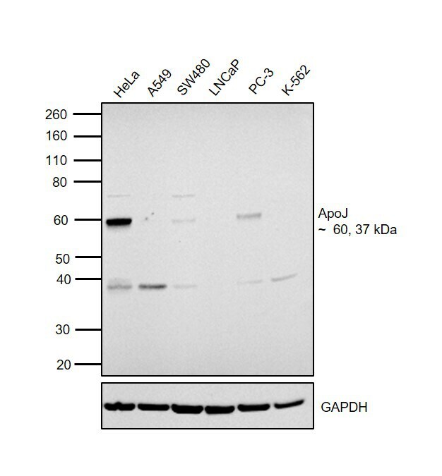
- Western blot was performed using Anti-Apolipoprotein J Mouse Monoclonal Antibody (Product # MA1-19385) and 37, 60 kDa bands corresponding to Apolipoprotein J was observed across cell lines tested except LNCaP. An additional band at ~ 38 kDa was observed across cell lines tested. Whole cell extracts (30 µg lysate) of HeLa (Lane 1), A549 (Lane 2), SW480 (Lane 3), LNCap (Lane 4), PC-3 (Lane 5) and K-562 (Lane 6) were electrophoresed using NuPAGE™ 4-12% Bis-Tris Protein Gel (Product # NP0322BOX). Resolved proteins were then transferred onto a nitrocellulose membrane (Product # IB23001) by iBlot® 2 Dry Blotting System (Product # IB21001). The blot was probed with the primary antibody (1 µg/mL) and detected by chemiluminescence with Goat anti-Mouse IgG (H+L) Superclonal™ Recombinant Secondary Antibody, HRP (Product # A28177, 1:4000 dilution) using the iBright FL 1000 (Product # A32752). Chemiluminescent detection was performed using Novex® ECL Chemiluminescent Substrate Reagent Kit (Product # WP20005).
Supportive validation
- Submitted by
- Invitrogen Antibodies (provider)
- Main image

- Experimental details
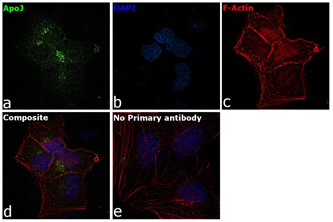
- Immunofluorescence analysis of Apolipoprotein J was performed using HeLa cells. The cells were fixed with 4% paraformaldehyde for 10 minutes, permeabilized with 0.1% Triton™ X-100 for 15 minutes, and blocked with 2% BSA for 1 hour at room temperature. The cells were labeled with Apolipoprotein J Mouse Monoclonal Antibody (Product # MA1-19385) at 5 µg/mL in 0.1% BSA and incubated overnight at 4 degree and then labeled with Goat anti-Mouse IgG (H+L) Highly Cross-Adsorbed Secondary Antibody, Alexa Fluor Plus 488 (Product # A32723) at a dilution of 1:2000 for 45 minutes at room temperature (Panel a: green) in HeLa cells. Nuclei (Panel b: blue) were stained with ProLong™ Diamond Antifade Mountant with DAPI (Product # P36962). F-actin (Panel c: red) was stained with Rhodamine Phalloidin (Product # R415, 1:300). Panel d represents the merged image of HeLa cells showing cytoplasmic and nuclear localization. Panel e represents control cells with no primary antibody to assess background. The images were captured at 60X magnification.