 Explore
 Validate
 Learn
LearnMAB29372-100
antibody from R&D Systems
Targeting: CLU
APOJ, CLI, CLU1, CLU2, KUB1, SGP-2, SP-40, TRPM-2
 ELISA
ELISA Immunohistochemistry
Antibody data
- Antibody Data
- Antigen structure
- References [0]
- Comments [0]
- Validations
- Immunohistochemistry [2]
Submit
Validation data
Reference
Comment
Report error
- Product number
- MAB29372-100 - Provider product page

- Provider
- R&D Systems
- Product name
- Human Clusterin Antibody
- Antibody type
- Monoclonal
- Description
- Protein A or G purified from hybridoma culture supernatant. Detects recombinant human Clusterin in ELISAs.
- Reactivity
- Human
- Host
- Mouse
- Conjugate
- Unconjugated
- Antigen sequence
NP_001822- Isotype
- IgG
- Antibody clone number
- 350270
- Vial size
- 100 ug
- Storage
- Use a manual defrost freezer and avoid repeated freeze-thaw cycles. 12 months from date of receipt, -20 to -70 °C as supplied. 1 month, 2 to 8 °C under sterile conditions after reconstitution. 6 months, -20 to -70 °C under sterile conditions after reconstitution.
No comments: Submit comment
Supportive validation
- Submitted by
- R&D Systems (provider)
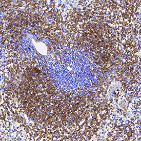
- Main image

- Experimental details
- Clusterin in Mouse Spleen. Clusterin was detected in immersion fixed frozen sections of mouse spleen using Mouse Anti-Human Clusterin Monoclonal Antibody (Catalog # MAB29372) at 5 µg/mL for 1 hour at room temperature followed by incubation with the Anti-Mouse IgG VisUCyte™ HRP Polymer Antibody (Catalog # VC001). Tissue was stained using DAB (brown) and counterstained with hematoxylin (blue). Specific staining was localized to cytoplasm. View our protocol for IHC Staining with VisUCyte HRP Polymer Detection Reagents.
- Submitted by
- R&D Systems (provider)
- Main image

- Experimental details
- Clusterin in Human Liver. Clusterin was detected in immersion fixed paraffin-embedded sections of human liver using Mouse Anti-Human Clusterin Monoclonal Antibody (Catalog # MAB29372) at 1 µg/mL for 1 hour at room temperature followed by incubation with the Anti-Mouse IgG VisUCyte™ HRP Polymer Antibody (Catalog # VC001). Tissue was stained using DAB (brown) and counterstained with hematoxylin (blue). Specific staining was localized to plasma membrane and cytoplasm. View our protocol for IHC Staining with VisUCyte HRP Polymer Detection Reagents.
