 Explore
 Validate
 Learn
Learn Western blot
 Immunocytochemistry
Antibody data
- Antibody Data
- Antigen structure
- References [0]
- Comments [0]
- Validations
- Immunocytochemistry [3]
- Immunoprecipitation [1]
- Immunohistochemistry [4]
- Chromatin Immunoprecipitation [3]
- Other assay [1]
Submit
Validation data
Reference
Comment
Report error
- Product number
- PA5-31828 - Provider product page

- Provider
- Invitrogen Antibodies
- Product name
- KDM6A Polyclonal Antibody
- Antibody type
- Polyclonal
- Antigen
- Recombinant full-length protein
- Description
- Recommended positive controls: SK-N-SH, IMR32, SK-N-AS. Predicted reactivity: Mouse (93%), Xenopus laevis (82%), Dog (86%), Bovine (98%). Store product as a concentrated solution. Centrifuge briefly prior to opening the vial.
- Reactivity
- Human, Mouse
- Host
- Rabbit
- Isotype
- IgG
- Vial size
- 100 μL
- Concentration
- 0.78 mg/mL
- Storage
- Store at 4°C short term. For long term storage, store at -20°C, avoiding freeze/thaw cycles.
No comments: Submit comment
Supportive validation
- Submitted by
- Invitrogen Antibodies (provider)
- Main image
- Experimental details
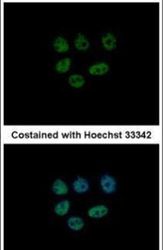
- Immunofluorescent analysis of KDM6A in paraformaldehyde-fixed MCF-7 cells using a KDM6A polyclonal antibody (Product # PA5-31828) at a 1:500 dilution.

- Submitted by
- Invitrogen Antibodies (provider)
- Main image

- Experimental details
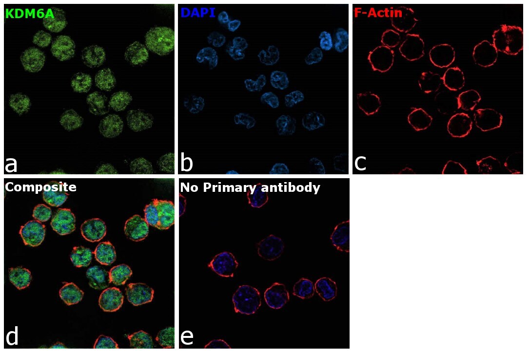
- Immunofluorescence analysis of KDM6A was performed using 70% confluent log phase K-562 cells. The cells were fixed with 4% paraformaldehyde for 10 minutes, permeabilized with 0.1% Triton™ X-100 for 15 minutes, and blocked with 2% BSA for 1 hour at room temperature. The cells were labeled with KDM6A Polyclonal Antibody (Product # PA5-31828) at 1:100 in 0.1% BSA, incubated at 4 degree celsius overnight and then labeled with Donkey anti-Rabbit IgG (H+L) Highly Cross-Adsorbed Secondary Antibody, Alexa Fluor Plus 488 (Product # A32790), (1:2000), for 45 minutes at room temperature (Panel a: Green). Nuclei (Panel b:Blue) were stained with ProLong™ Diamond Antifade Mountant with DAPI (Product # P36962). F-actin (Panel c: Red) was stained with Rhodamine Phalloidin (Product # R415, 1:300). Panel d represents the merged image showing nuclear and cytosolic localization. Panel e represents control cells with no primary antibody to assess background. The images were captured at 60X with oil immersion magnification.

- Submitted by
- Invitrogen Antibodies (provider)
- Main image

- Experimental details
- Immunofluorescence analysis of KDM6A was performed using 70% confluent log phase K-562 cells. The cells were fixed with 4% paraformaldehyde for 10 minutes, permeabilized with 0.1% Triton™ X-100 for 15 minutes, and blocked with 2% BSA for 1 hour at room temperature. The cells were labeled with KDM6A Polyclonal Antibody (Product # PA5-31828) at 1:100 in 0.1% BSA, incubated at 4 degree celsius overnight and then labeled with Donkey anti-Rabbit IgG (H+L) Highly Cross-Adsorbed Secondary Antibody, Alexa Fluor Plus 488 (Product # A32790), (1:2000), for 45 minutes at room temperature (Panel a: Green). Nuclei (Panel b:Blue) were stained with ProLong™ Diamond Antifade Mountant with DAPI (Product # P36962). F-actin (Panel c: Red) was stained with Rhodamine Phalloidin (Product # R415, 1:300). Panel d represents the merged image showing nuclear and cytosolic localization. Panel e represents control cells with no primary antibody to assess background. The images were captured at 60X with oil immersion magnification.
Supportive validation
- Submitted by
- Invitrogen Antibodies (provider)
- Main image

- Experimental details
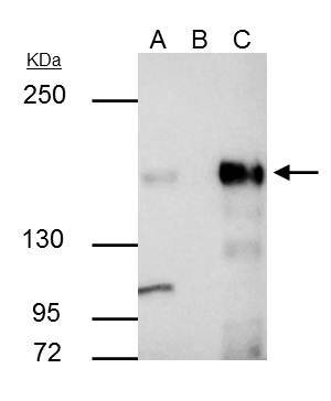
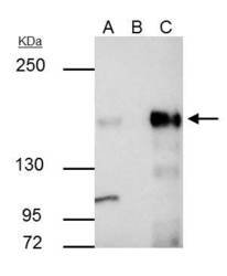
- KDM6A Polyclonal Antibody immunoprecipitates KDM6A protein in IP experiments. IP samples: SK-N-SH whole cell extract. A. 30 µg SK-N-SH whole cell extract. B. Control with 4 µg of preimmune Rabbit IgG. C. Immunoprecipitation of KDM6A protein by 4 µg KDM6A Polyclonal Antibody (Product # PA5-31828). 5 % SDS-PAGE. The immunoprecipitated KDM6A protein was detected by KDM6A Polyclonal Antibody (Product # PA5-31828) diluted at 1:500.
Supportive validation
- Submitted by
- Invitrogen Antibodies (provider)
- Main image
- Experimental details
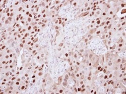
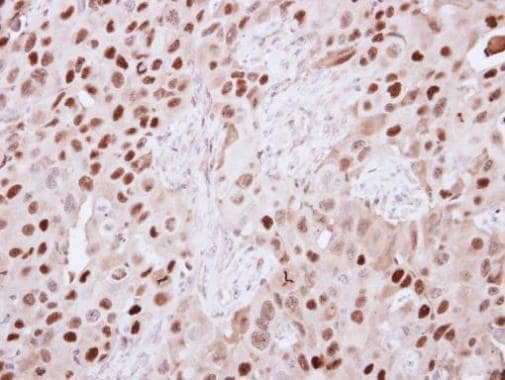
- Immunohistochemical analysis of paraffin-embedded H441 xenograft, using KDM6A (Product # PA5-31828) antibody at 1:250 dilution. Antigen Retrieval: EDTA based buffer, pH 8.0, 15 min.
- Submitted by
- Invitrogen Antibodies (provider)
- Main image
- Experimental details
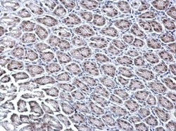
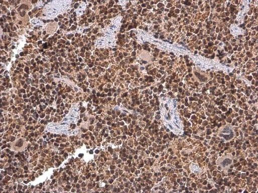
- KDM6A Polyclonal Antibody detects KDM6A protein at nucleus on mouse duodenum by immunohistochemical analysis. Sample: Paraffin-embedded mouse duodenum. KDM6A Polyclonal Antibody (Product # PA5-31828) dilution: 1:1,000. Antigen Retrieval: EDTA based buffer, pH 8.0, 15 min.
- Submitted by
- Invitrogen Antibodies (provider)
- Main image
- Experimental details
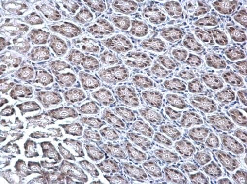
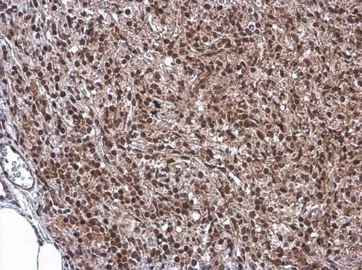
- Immunohistochemistry (Paraffin) analysis of KDM6A was performed in paraffin-embedded mouse lymph node tissue using KDM6A Polyclonal Antibody (Product # PA5-31828) at a dilution of 1:500. Antigen Retrieval: Citrate buffer, pH 6.0, 15 min.
- Submitted by
- Invitrogen Antibodies (provider)
- Main image
- Experimental details
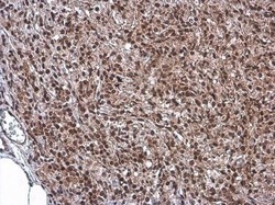
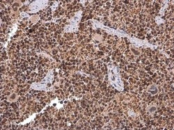
- Immunohistochemistry (Paraffin) analysis of KDM6A was performed in paraffin-embedded mouse spleen tissue using KDM6A Polyclonal Antibody (Product # PA5-31828) at a dilution of 1:500. Antigen Retrieval: Citrate buffer, pH 6.0, 15 min.
Supportive validation
- Submitted by
- Invitrogen Antibodies (provider)
- Main image
- Experimental details
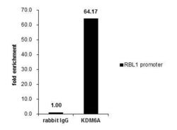
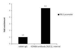
- Cross-linked ChIP was performed with HeLa chromatin extract and 5 µg of either control rabbit IgG or a KDM6A polyclonal antibody (Product # PA5-31828). The precipitated DNA was detected by PCR with primer set targeting to RBL1 promoter.

- Submitted by
- Invitrogen Antibodies (provider)
- Main image

- Experimental details
- ChIP assay analysis of KDM6A in HeLa chromatin extracts using 5 µg of either normal rabbit IgG or KDM6A Polyclonal Antibody (Product # PA5-31828). The precipitated DNA was detected by PCR with primer set targeting to RBL1 promoter.

- Submitted by
- Invitrogen Antibodies (provider)
- Main image
- Experimental details
- ChIP assay analysis of KDM6A in HeLa chromatin extracts using 5 µg of either normal rabbit IgG or KDM6A Polyclonal Antibody (Product # PA5-31828). The precipitated DNA was detected by PCR with primer set targeting to RBL1 promoter.

Supportive validation
- Submitted by
- Invitrogen Antibodies (provider)
- Main image
- Experimental details
- KDM6A Polyclonal Antibody immunoprecipitates KDM6A protein in IP experiments. IP samples: SK-N-SH whole cell extract. A. 30 µg SK-N-SH whole cell extract. B. Control with 4 µg of preimmune Rabbit IgG. C. Immunoprecipitation of KDM6A protein by 4 µg KDM6A Polyclonal Antibody (Product # PA5-31828). 5 % SDS-PAGE. The immunoprecipitated KDM6A protein was detected by KDM6A Polyclonal Antibody (Product # PA5-31828) diluted at 1:500.
