Explore
 Validate
 Learn
Learn Western blot
Antibody data
- Antibody Data
- Antigen structure
- References [1]
- Comments [0]
- Validations
- Western blot [1]
- Immunocytochemistry [1]
- Other assay [1]
Submit
Validation data
Reference
Comment
Report error
- Product number
- PA5-30403 - Provider product page

- Provider
- Invitrogen Antibodies
- Product name
- TCP-1 theta Polyclonal Antibody
- Antibody type
- Polyclonal
- Antigen
- Recombinant full-length protein
- Description
- Recommended positive controls: 293T, A431, Jurkat, Raji. Predicted reactivity: Mouse (97%), Rat (98%), Zebrafish (84%), Xenopus laevis (87%), Chicken (95%), Rhesus Monkey (99%), Chimpanzee (98%), Bovine (98%). Store product as a concentrated solution. Centrifuge briefly prior to opening the vial.
- Reactivity
- Human
- Host
- Rabbit
- Isotype
- IgG
- Vial size
- 100 μL
- Concentration
- 0.99 mg/mL
- Storage
- Store at 4°C short term. For long term storage, store at -20°C, avoiding freeze/thaw cycles.
Submitted references ARP-T1-associated Bazex-Dupré-Christol syndrome is an inherited basal cell cancer with ciliary defects characteristic of ciliopathies.
Park HS, Papanastasi E, Blanchard G, Chiticariu E, Bachmann D, Plomann M, Morice-Picard F, Vabres P, Smahi A, Huber M, Pich C, Hohl D
Communications biology 2021 May 10;4(1):544
Communications biology 2021 May 10;4(1):544
No comments: Submit comment
Supportive validation
- Submitted by
- Invitrogen Antibodies (provider)
- Main image

- Experimental details
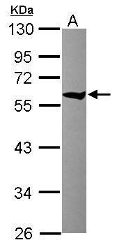
- Western Blot using TCP-1 theta Polyclonal Antibody (Product # PA5-30403). Sample (30 µg of whole cell lysate). Lane A: Raji. 10% SDS PAGE. TCP-1 theta Polyclonal Antibody (Product # PA5-30403) diluted at 1:10,000.
Supportive validation
- Submitted by
- Invitrogen Antibodies (provider)
- Main image
- Experimental details
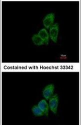
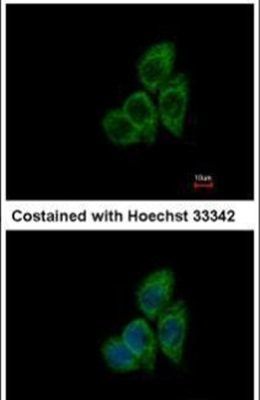
- Immunofluorescent analysis of TCP1 theta in methanol-fixed A431 cells using a TCP1 theta polyclonal antibody (Product # PA5-30403) at a 1:500 dilution.
Supportive validation
- Submitted by
- Invitrogen Antibodies (provider)
- Main image
- Experimental details
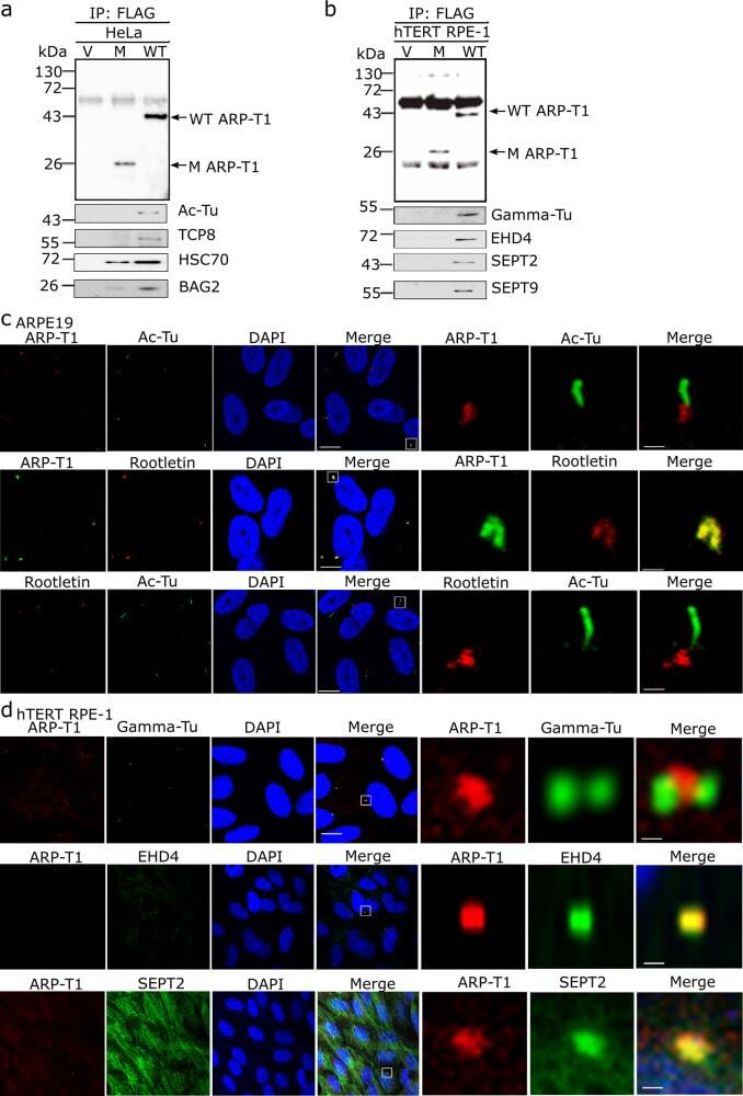
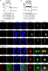
- Fig. 3 ARP-T1 interacts with proteins involved in ciliary machinery. a , b HeLa ( a ) and hTERT-RPE1 ( b ) cells were transduced with lentiviral vectors, empty vector (V), ACTRT1 mutant (M) and ACTRT1 WT (WT), and immunoprecipitated (IP) with anti-FLAG monoclonal antibody M2-conjugated agarose, and analyzed by immunoblot with indicated antisera. c Immunofluorescence stainings of ARP-T1, acetylated-tubulin and rootletin in 35 days of serum-starved ARPE19 cells. Nuclei are stained with DAPI. Scale bar, 5 um. Higher magnifications of the boxed area are shown on right three panels. Scale bar, 1 um. d Immunofluorescence staining of ARP-T1, gamma-tubulin, EHD4, and septin 2 in 48 h of serum-starved hTERT-RPE1 cells. Nuclei are stained with DAPI. Scale bar, 5 um. Higher magnifications of the boxed area are shown on the right three panels. Scale bar, 1 um.