Explore
 Validate
 Learn
Learn Western blot
 Immunocytochemistry
Antibody data
- Antibody Data
- Antigen structure
- References [0]
- Comments [0]
- Validations
- Immunocytochemistry [1]
- Immunohistochemistry [1]
- Flow cytometry [2]
Submit
Validation data
Reference
Comment
Report error
- Product number
- PA5-25405 - Provider product page

- Provider
- Invitrogen Antibodies
- Product name
- USP17 Polyclonal Antibody
- Antibody type
- Polyclonal
- Antigen
- Synthetic peptide
- Reactivity
- Human
- Host
- Rabbit
- Isotype
- IgG
- Vial size
- 400 μL
- Concentration
- 0.5 mg/mL
- Storage
- Store at 4°C short term. For long term storage, store at -20°C, avoiding freeze/thaw cycles.
No comments: Submit comment
Supportive validation
- Submitted by
- Invitrogen Antibodies (provider)
- Main image
- Experimental details
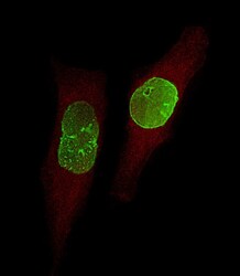
- Immunocytochemistry analysis of USP17 in U-251 MG cells. Samples were incubated with USP17 polyclonal antibody (Product # PA5-25405) using a dilution of 1:25 followed by Dylight® 488-conjugated goat anti-Rabbit IgG at a dilution of 1:200 (green). Cells were 4% paraformaldehyde-fixed, 0. 1% Triton X-100 permeabilized. Immunofluorescence image showing Nucleus staining on U-251 MG cell line. Cytoplasmic actin is detected with Dylight® 554 Phalloidin(red). The nuclear counter stain is DAPI (blue).

Supportive validation
- Submitted by
- Invitrogen Antibodies (provider)
- Main image
- Experimental details
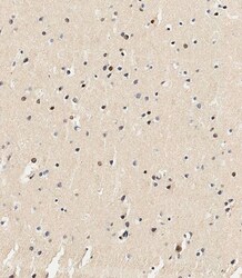
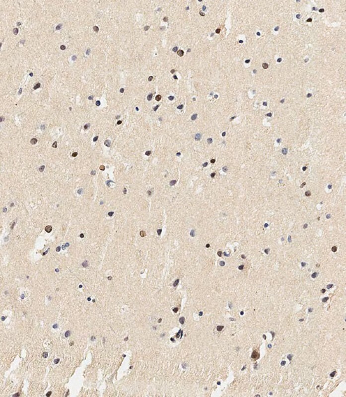
- Immunohistochemistry analysis of USP17 in paraffin-embedded human brain tissue. Samples were incubated with USP17 polyclonal antibody (Product # PA5-25405) using a dilution of 1:500 for 1 hour at room temperature followed by an undiluted biotinylated CRF Anti-Polyvalent HRP Polymer antibody. Tissue was fixed with formaldehyde at room temperature, antigen retrieval was by heat mediation with a EDTA buffer (pH 9.0).
Supportive validation
- Submitted by
- Invitrogen Antibodies (provider)
- Main image
- Experimental details
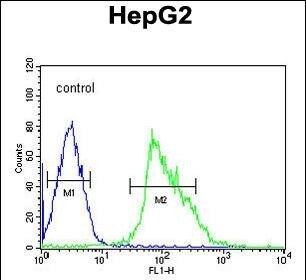
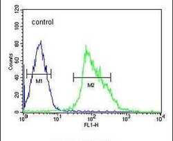
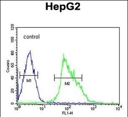
- Flow cytometry analysis of HepG2 cells using a USP17 polyclonal antibody (Product # PA5-25405) (right) compared to a negative control cell (left) at a dilution of 1:10-50, followed by a FITC-conjugated goat anti-rabbit antibody
- Submitted by
- Invitrogen Antibodies (provider)
- Main image
- Experimental details
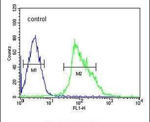
- Flow cytometry of USP17 in HepG2 cells (right histogram). Samples were incubated with USP17 polyclonal antibody (Product # PA5-25405) followed by FITC-conjugated goat-anti-rabbit secondary antibody. Negative control cell (left histogram).