 Explore
 Validate
 Learn
Learn Western blot
 Immunohistochemistry
Antibody data
- Antibody Data
- Antigen structure
- References [2]
- Comments [0]
- Validations
- Western blot [1]
- Immunocytochemistry [4]
- Chromatin Immunoprecipitation [2]
Submit
Validation data
Reference
Comment
Report error
- Product number
- MA5-17078 - Provider product page

- Provider
- Invitrogen Antibodies
- Product name
- FOXO1 Monoclonal Antibody (3B6)
- Antibody type
- Monoclonal
- Antigen
- Purifed from natural sources
- Description
- MA5-17078 targets FOXO1 in ChIP, IHC, IF, and WB applications and shows reactivity with Human and Mouse samples. The MA5-17078 immunogen is purified recombinant fragment of human FOXO1 expressed in E. Coli. MA5-17078 detects FOXO1 which has a predicted molecular weight of approximately 69.7kDa.
- Reactivity
- Human, Mouse
- Host
- Mouse
- Isotype
- IgG
- Antibody clone number
- 3B6
- Vial size
- 100 μg
- Concentration
- 1 mg/mL
- Storage
- Store at 4°C short term. For long term storage, store at -20°C, avoiding freeze/thaw cycles.
Submitted references Hydrogen sulphide mitigates homocysteine-induced apoptosis and matrix remodelling in mesangial cells through Akt/FOXO1 signalling cascade.
3D Culture Method for Alzheimer's Disease Modeling Reveals Interleukin-4 Rescues Aβ42-Induced Loss of Human Neural Stem Cell Plasticity.
Majumder S, Ren L, Pushpakumar S, Sen U
Cellular signalling 2019 Sep;61:66-77
Cellular signalling 2019 Sep;61:66-77
3D Culture Method for Alzheimer's Disease Modeling Reveals Interleukin-4 Rescues Aβ42-Induced Loss of Human Neural Stem Cell Plasticity.
Papadimitriou C, Celikkaya H, Cosacak MI, Mashkaryan V, Bray L, Bhattarai P, Brandt K, Hollak H, Chen X, He S, Antos CL, Lin W, Thomas AK, Dahl A, Kurth T, Friedrichs J, Zhang Y, Freudenberg U, Werner C, Kizil C
Developmental cell 2018 Jul 2;46(1):85-101.e8
Developmental cell 2018 Jul 2;46(1):85-101.e8
No comments: Submit comment
Supportive validation
- Submitted by
- Invitrogen Antibodies (provider)
- Main image
- Experimental details
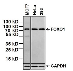
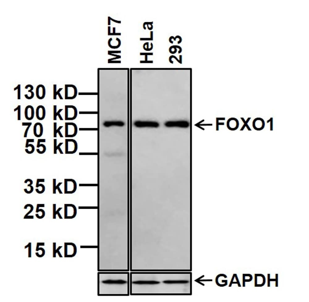
- Western blot analysis of FOXO1 was performed by loading 20 µg of MCF7 whole cell lysate and 7 µL of PageRuler Plus Prestained Protein Ladder (Product # 26619) per well onto a 4-20% Tris-Glycine polyacrylamide gel (Product # WT4202BX10). Proteins were transferred to a nitrocellulose membrane using the G2 Blotter (Product # 62288), and blocked with 5% BSA in TBST (Product # 37520) for 1 hour at room temperature. FOXO1 was detected at ~72 kDa using a FOXO1 mouse monoclonal antibody (Product # MA5-17078) at a dilution of 1:500 in blocking buffer overnight at 4°C on a rocking platform, followed by a Goat anti-Mouse IgG (H+L) Superclonal™ Secondary Antibody, HRP conjugate (Product # A28177) at a dilution of 1:1000 for at least 30 minutes at room temperature. dy, HRP conjugate (Product # A28177) at a dilution of 1:2000 for at least 30 minutes at room temperature. Chemiluminescent detection was performed using SuperSignal West Pico (Product # 34078).
Supportive validation
- Submitted by
- Invitrogen Antibodies (provider)
- Main image
- Experimental details
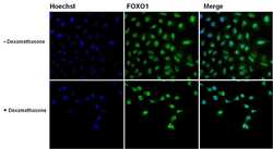
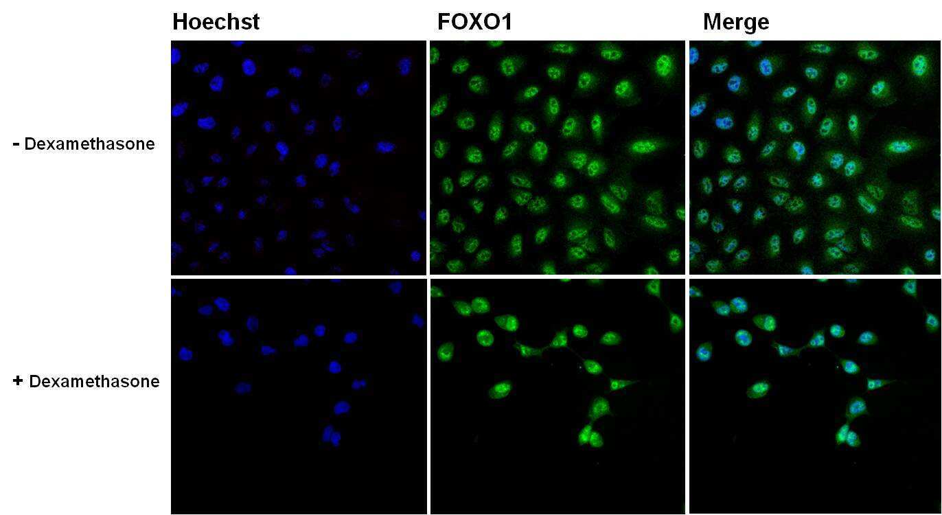
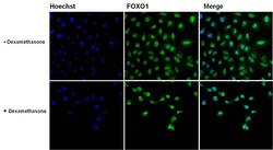
- Immunofluorescent analysis of FOXO1 (green) in untreated and 1uM 20 hours Dexamethasone treated A549 cells. The cells were fixed with 4% paraformaldehyde for 15 minutes at -20c, permeabilized with 0.1% Triton X-100 for 15 minutes, and blocked with 3% BSA for 30 minutes at room temperature. Cells were stained with a FOXO1 mouse monoclonal antibody (Product # MA5-17078) at a concentration of 5 µg/mL in blocking buffer for 1 hour at room temperature, and then incubated with a Goat anti-Mouse IgG (H+L) Secondary Antibody, Alexa Fluor Plus 488 conjugate (Product # A32731) at a dilution of 1:500 for at least 30 minutes at a room temperature in the dark (green). Nuclei (blue) were stained with Hoechst 33342 (Product # 62249). Images were taken on a Thermo Scientific ToxInsight Instrument at 20X magnification.
- Submitted by
- Invitrogen Antibodies (provider)
- Main image
- Experimental details
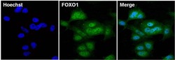
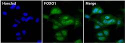
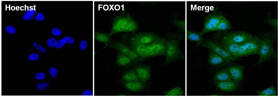
- Immunofluorescent analysis of FOXO1 (green) in MCF7 cells. The cells were fixed with 4% paraformaldehyde for 15 minutes at -20c, permeabilized with 0.1% Triton X-100 for 15 minutes, and blocked with 3% BSA for 30 minutes at room temperature. Cells were stained with a FOXO1 mouse monoclonal antibody (Product # MA5-17078) at a concentration of 5 µg/mL in blocking buffer for 1 hour at room temperature, and then incubated with a Goat anti-Mouse IgG (H+L) Secondary Antibody, Alexa Fluor Plus 488 conjugate (Product # A32731) at a dilution of 1:500 for at least 30 minutes at a room temperature in the dark (green). Nuclei (blue) were stained with Hoechst 33342 (Product # 62249). Images were taken on a Thermo Scientific ToxInsight Instrument at 20X magnification.

- Submitted by
- Invitrogen Antibodies (provider)
- Main image
- Experimental details
- Immunofluorescent analysis of FOXO1 (green) in untreated and 1uM 20 hours Dexamethasone treated A549 cells. The cells were fixed with 4% paraformaldehyde for 15 minutes at -20c, permeabilized with 0.1% Triton X-100 for 15 minutes, and blocked with 3% BSA for 30 minutes at room temperature. Cells were stained with a FOXO1 mouse monoclonal antibody (Product # MA5-17078) at a concentration of 5 µg/mL in blocking buffer for 1 hour at room temperature, and then incubated with a Goat anti-Mouse IgG (H+L) Secondary Antibody, Alexa Fluor Plus 488 conjugate (Product # A32731) at a dilution of 1:500 for at least 30 minutes at a room temperature in the dark (green). Nuclei (blue) were stained with Hoechst 33342 (Product # 62249). Images were taken on a Thermo Scientific ToxInsight Instrument at 20X magnification.

- Submitted by
- Invitrogen Antibodies (provider)
- Main image
- Experimental details
- Immunofluorescent analysis of FOXO1 (green) in MCF7 cells. The cells were fixed with 4% paraformaldehyde for 15 minutes at -20c, permeabilized with 0.1% Triton X-100 for 15 minutes, and blocked with 3% BSA for 30 minutes at room temperature. Cells were stained with a FOXO1 mouse monoclonal antibody (Product # MA5-17078) at a concentration of 5 µg/mL in blocking buffer for 1 hour at room temperature, and then incubated with a Goat anti-Mouse IgG (H+L) Secondary Antibody, Alexa Fluor Plus 488 conjugate (Product # A32731) at a dilution of 1:500 for at least 30 minutes at a room temperature in the dark (green). Nuclei (blue) were stained with Hoechst 33342 (Product # 62249). Images were taken on a Thermo Scientific ToxInsight Instrument at 20X magnification.
Supportive validation
- Submitted by
- Invitrogen Antibodies (provider)
- Main image
- Experimental details
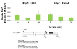
- Multiplex microplate Matrix ChIP has been described in detail (http://www.ncbi.nlm.nih.gov/pubmed/25959381). Briefly HTC116 cells were starved followed by addition of serum and samples of cells were cross-linked with formaldehyde after the time points indicated on the x-axis (0, 5 and 30 min). Chromatin was sheared using a Bioruptor and ChIP assays were performed using protein A-coated 96-well polypropylene microplates with 1uL/100uL well volume of FOXO1 monoclonal antibody (Product # MA5-17078). Quantitative real-time PCRs were performed in quadruplicate using 1 to 2uL of DNA with primers to -15kb downstream of Egr1 and exon 1 of Erg1. PCR calibration curves were generated for each primer pair from a dilution series of total human genomic DNA. The PCR primer efficiency curve was fit to cycle threshold (Ct) versus log [genomic DNA concentration] by using an r2 best fit. DNA concentration values for each ChIP and input DNA sample were calculated from their respective average Ct values. Final results are expressed as fraction of input DNA. Schematic representations of Erg1 loci is shown where boxes represent exons (black boxes = translated regions, white boxes = untranslated regions), the zigzag line represents an intron, and the straight line represents upstream sequence. Regions amplified by the primers are represented by black bars. Data courtesy of Dr. Karol Bomsztyk’s laboratory.

- Submitted by
- Invitrogen Antibodies (provider)
- Main image

- Experimental details
- Multiplex microplate Matrix ChIP has been described in detail (http://www.ncbi.nlm.nih.gov/pubmed/25959381). Briefly HTC116 cells were starved followed by addition of serum and samples of cells were cross-linked with formaldehyde after the time points indicated on the x-axis (0, 5 and 30 min). Chromatin was sheared using a Bioruptor and ChIP assays were performed using protein A-coated 96-well polypropylene microplates with 1uL/100uL well volume of FOXO1 monoclonal antibody (Product # MA5-17078). Quantitative real-time PCRs were performed in quadruplicate using 1 to 2uL of DNA with primers to -15kb downstream of Egr1 and exon 1 of Erg1. PCR calibration curves were generated for each primer pair from a dilution series of total human genomic DNA. The PCR primer efficiency curve was fit to cycle threshold (Ct) versus log [genomic DNA concentration] by using an r2 best fit. DNA concentration values for each ChIP and input DNA sample were calculated from their respective average Ct values. Final results are expressed as fraction of input DNA. Schematic representations of Erg1 loci is shown where boxes represent exons (black boxes = translated regions, white boxes = untranslated regions), the zigzag line represents an intron, and the straight line represents upstream sequence. Regions amplified by the primers are represented by black bars. Data courtesy of Dr. Karol Bomsztyk’s laboratory.
