 Explore
 Validate
 Learn
Learn Western blot
 Immunocytochemistry
Antibody data
- Antibody Data
- Antigen structure
- References [2]
- Comments [0]
- Validations
- Western blot [3]
- Immunohistochemistry [2]
- Flow cytometry [4]
Submit
Validation data
Reference
Comment
Report error
- Product number
- NBP2-31376 - Provider product page

- Provider
- Novus Biologicals
- Product name
- Mouse Monoclonal FoxO1/FKHR Antibody
- Antibody type
- Monoclonal
- Description
- Protein G purified.
- Reactivity
- Human, Rat
- Host
- Mouse
- Isotype
- IgG
- Vial size
- 0.1 mg
- Concentration
- 1.0 mg/ml
- Storage
- Store at 4C short term. Aliquot and store at -20C long term. Avoid freeze-thaw cycles.
Submitted references Regular exercise potentiates energetically expensive hepatic de novo lipogenesis during early weight regain.
Histone H3 lysine 9 acetylation is downregulated in GDM Placentas and Calcitriol supplementation enhanced this effect.
Presby DM, Checkley LA, Jackman MR, Higgins JA, Jones KL, Giles ED, Houck JA, Webb PG, Steig AJ, Johnson GC, Rudolph MC, MacLean PS
American journal of physiology. Regulatory, integrative and comparative physiology 2019 Nov 1;317(5):R684-R695
American journal of physiology. Regulatory, integrative and comparative physiology 2019 Nov 1;317(5):R684-R695
Histone H3 lysine 9 acetylation is downregulated in GDM Placentas and Calcitriol supplementation enhanced this effect.
Hepp P, Hutter S, Knabl J, Hofmann S, Kuhn C, Mahner S, Jeschke U
International journal of molecular sciences 2018 Dec 14;19(12)
International journal of molecular sciences 2018 Dec 14;19(12)
No comments: Submit comment
Supportive validation
- Submitted by
- Novus Biologicals (provider)
- Main image

- Experimental details
- Simple Western: FoxO1/FKHR Antibody (83N7F8) [NBP2-31376] - Lane view shows a specific band for FoxO1/FKHR in 0.5 mg/ml of Hek293 lysate. This experiment was performed under reducing conditions using the 12-230 kDa separation system.

- Submitted by
- Novus Biologicals (provider)
- Main image
- Experimental details
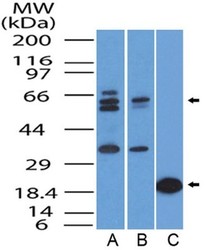
- Western Blot: FoxO1/FKHR Antibody (83N7F8) [NBP2-31376] - Detection of FOXO1A / FOXO1 protein in (A) Hela cells lysate, (B) Jurkat cell lysate, and (C) partial recombinant protein by using FOXO1A antibody at a concentration of 0.5 ug/ml for lysate and 0.05 ug/ml for the recombinant protein.

- Submitted by
- Novus Biologicals (provider)
- Main image

- Experimental details
- Western Blot: FoxO1/FKHR Antibody (83N7F8) [NBP2-31376] - Analysis of (A) Ntera2 cell lysate, (B) HEK293 cell lysate, and (C) human placenta tissue using FOXO1A antibody at a concentration of 2 ug/ml.

Supportive validation
- Submitted by
- Novus Biologicals (provider)
- Main image

- Experimental details
- Immunohistochemistry-Paraffin: FoxO1/FKHR Antibody (83N7F8) [NBP2-31376] - Analysis of FOXO1A protein in a section of normal human uterus by using FOXO1A antibody at a concentration of 5 ug/ml. The columnar cells of straight non-convoluted glands in uterous showed specific staining for FOXO1A in the cellular cytoplasm as well as nuclei, whereas the stromal cells showed mild cytoplasmic staining only.

- Submitted by
- Novus Biologicals (provider)
- Main image

- Experimental details
- Immunohistochemistry-Paraffin: FoxO1/FKHR Antibody (83N7F8) [NBP2-31376] - Analysis of FOXO1A protein in a section of normal human kidney by using FOXO1A antibody at a concentration of 5 ug/ml. Cytoplasmic and nuclear positivity was observed in the cells of various tubules/ducts of renal medulla.

Supportive validation
- Submitted by
- Novus Biologicals (provider)
- Main image

- Experimental details
- Flow Cytometry: FoxO1/FKHR Antibody (83N7F8) [NBP2-31376] - Analysis using the Alexa Fluor 488 conjugated FoxO1 antibody, NBP2-31376AF488 (blue) and fluorescence minus one control (red). Staining of FoxO1 in primary CD4+ T-cells isolated from human peripheral blood using 1uL of antibody per 1X10e5 cells. Image from verified customer review.

- Submitted by
- Novus Biologicals (provider)
- Main image

- Experimental details
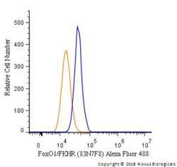
- Flow Cytometry: FoxO1/FKHR Antibody (83N7F8) [NBP2-31376] - Using the Alexa Fluor 488 direct conjugate An intracellular stain was performed on Daudi cells with FoxO1/FKHR (83N7F8) antibody NBP2-31376AF488 (blue) and a matched isotype control NBP2-27231AF488 (orange). Cells were fixed with 4% PFA and then permeablized with 0.1% saponin. Cells were incubated in an antibody dilution of 5 ug/mL for 30 minutes at room temperature. Both antibodies were conjugated to Alexa Fluor 488.

- Submitted by
- Novus Biologicals (provider)
- Main image
- Experimental details
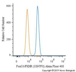
- Flow Cytometry: FoxO1/FKHR Antibody (83N7F8) [NBP2-31376] - An intracellular stain was performed on U2-OS cells with FoxO1/FKHR [83N7F8] Antibody NBP2-31376AF488 (blue) and a matched isotype control (orange). Cells were fixed with 4% PFA and then permeabilized with 0.1% saponin. Cells were incubated in an antibody dilution of 10 ug/mL for 30 minutes at room temperature. Both antibodies were conjugated to Alexa Fluor 488.

- Submitted by
- Novus Biologicals (provider)
- Main image
- Experimental details
- Flow Cytometry: FoxO1/FKHR Antibody (83N7F8) [NBP2-31376] - An intracellular stain was performed on RH-30 cells with FoxO1/FKHR Antibody [83N7F8] NBP2-31376AF488 (blue) and a matched isotype control (orange). Cells were fixed with 4% PFA and then permeabilized with 0.1% saponin. Cells were incubated in an antibody dilution of 5 ug/mL for 30 minutes at room temperature. Both antibodies were conjugated to Alexa Fluor 488.
