 Explore
 Validate
 Learn
Learn Western blot
 Immunocytochemistry
 Immunoprecipitation
Antibody data
- Antibody Data
- Antigen structure
- References [2]
- Comments [0]
- Validations
- Immunocytochemistry [2]
- Immunohistochemistry [1]
- Other assay [1]
Submit
Validation data
Reference
Comment
Report error
- Product number
- MA1-91639 - Provider product page

- Provider
- Invitrogen Antibodies
- Product name
- HSPA9 Monoclonal Antibody (30A5)
- Antibody type
- Monoclonal
- Antigen
- Other
- Description
- Western blot detects a band ~75 kDa. A suggested positive control is HeLa cell lysate (heat shocked).
- Reactivity
- Human, Mouse, Rat, Bovine, Canine, Guinea Pig, Hamster, Porcine, Rabbit, Xenopus
- Host
- Mouse
- Isotype
- IgG
- Antibody clone number
- 30A5
- Vial size
- 25 μL
- Concentration
- 1 mg/mL
- Storage
- Store at 4°C short term. For long term storage, store at -20°C, avoiding freeze/thaw cycles.
Submitted references A Proteomic Study Suggests Stress Granules as New Potential Actors in Radiation-Induced Bystander Effects.
75-kDa glucose-regulated protein (GRP75) is a novel molecular signature for heat stress response in avian species.
Tudor M, Gilbert A, Lepleux C, Temelie M, Hem S, Armengaud J, Brotin E, Haghdoost S, Savu D, Chevalier F
International journal of molecular sciences 2021 Jul 26;22(15)
International journal of molecular sciences 2021 Jul 26;22(15)
75-kDa glucose-regulated protein (GRP75) is a novel molecular signature for heat stress response in avian species.
Dhamad AE, Greene E, Sales M, Nguyen P, Beer L, Liyanage R, Dridi S
American journal of physiology. Cell physiology 2020 Feb 1;318(2):C289-C303
American journal of physiology. Cell physiology 2020 Feb 1;318(2):C289-C303
No comments: Submit comment
Supportive validation
- Submitted by
- Invitrogen Antibodies (provider)
- Main image
- Experimental details
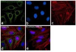
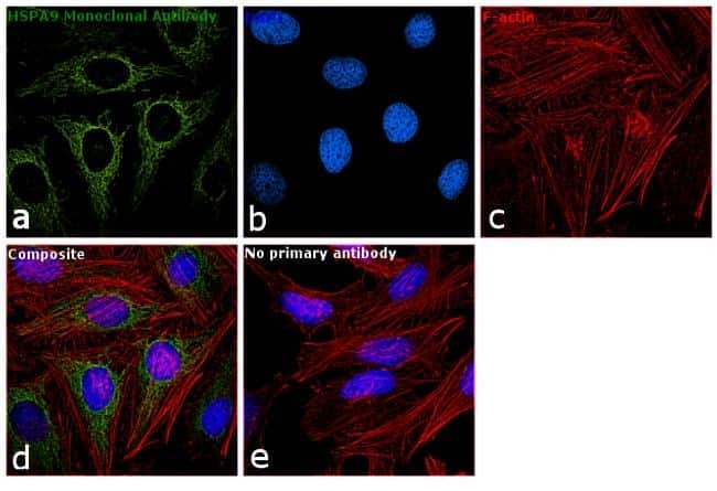
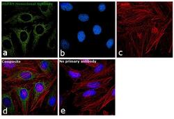
- Immunofluorescence analysis of HSPA9 was performed using 70% confluent log phase HeLa cells. The cells were fixed with 4% paraformaldehyde for 10 minutes, permeabilized with 0.1% Triton™ X-100 for 15 minutes, and blocked with 1% BSA for 1 hour at room temperature. The cells were labeled with HSPA9 Monoclonal Antibody (30A5) (Product # MA1-91639) at 1:100 dilution in 0.1% BSA, incubated at 4 degree Celsius overnight and then labeled with Goat anti-Mouse IgG (H+L) Superclonal™ Secondary Antibody, Alexa Fluor® 488 conjugate (Product # A28175) at a dilution of 1:2000 for 45 minutes at room temperature (Panel a: green). Nuclei (Panel b: blue) were stained with SlowFade® Gold Antifade Mountant with DAPI (Product # S36938). F-actin (Panel c: red) was stained with Rhodamine Phalloidin (Product # R415, 1:300). Panel d represents the merged image showing mitochondrial localization. Panel e represents control cells with no primary antibody to assess background. The images were captured at 60X magnification.
- Submitted by
- Invitrogen Antibodies (provider)
- Main image
- Experimental details
- Immunofluorescence analysis of HSPA9 was performed using 70% confluent log phase HeLa cells. The cells were fixed with 4% paraformaldehyde for 10 minutes, permeabilized with 0.1% Triton™ X-100 for 15 minutes, and blocked with 1% BSA for 1 hour at room temperature. The cells were labeled with HSPA9 Monoclonal Antibody (30A5) (Product # MA1-91639) at 1:100 dilution in 0.1% BSA, incubated at 4 degree Celsius overnight and then labeled with Goat anti-Mouse IgG (H+L) Superclonal™ Secondary Antibody, Alexa Fluor® 488 conjugate (Product # A28175) at a dilution of 1:2000 for 45 minutes at room temperature (Panel a: green). Nuclei (Panel b: blue) were stained with SlowFade® Gold Antifade Mountant with DAPI (Product # S36938). F-actin (Panel c: red) was stained with Rhodamine Phalloidin (Product # R415, 1:300). Panel d represents the merged image showing mitochondrial localization. Panel e represents control cells with no primary antibody to assess background. The images were captured at 60X magnification.

Supportive validation
- Submitted by
- Invitrogen Antibodies (provider)
- Main image
- Experimental details
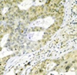
- Immunohistochemistry analysis of human breast cancer tissue immunohistochemically stained using HSPA9 Monoclonal Antibody (30A5) (Product # MA1-91639).

Supportive validation
- Submitted by
- Invitrogen Antibodies (provider)
- Main image
- Experimental details
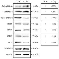
- Figure 5 Western blotting analysis of cyclophilin A, thioredoxin, alpha-enolase, RPLP0, HSC70, HSPA9, and CCT3 in a whole-cell extracts from T/C-28A2 bystander cells receiving the conditioned medium of low-dose irradiated chondrosarcoma cells (0.1 Gy) or non-irradiated chondrosarcoma cells (CTR).
