Explore
 Validate
 Learn
Learn Western blot
 Immunocytochemistry
Antibody data
- Antibody Data
- Antigen structure
- References [1]
- Comments [0]
- Validations
- Western blot [2]
- Immunohistochemistry [6]
- Flow cytometry [2]
Submit
Validation data
Reference
Comment
Report error
- Product number
- NBP1-47801 - Provider product page

- Provider
- Novus Biologicals
- Proper citation
- Novus Cat#NBP1-47801, RRID:AB_10010771
- Product name
- Mouse Monoclonal GRP75/HSPA9B/Mortalin Antibody
- Antibody type
- Monoclonal
- Description
- Affinity purified. This antibody is specific for Homo sapiens heat shock 70kDa protein 9 (mortalin) (HSPA9), nuclear gene encoding mitochondrial protein.
- Reactivity
- Human, Mouse, Rat, Canine, Simian
- Host
- Mouse
- Isotype
- IgG
- Vial size
- 0.1 ml
- Concentration
- 1 mg/ml
- Storage
- Store at -20C. Avoid freeze-thaw cycles.
Submitted references ERα36, a variant of estrogen receptor α, is predominantly localized in mitochondria of human uterine smooth muscle and leiomyoma cells.
Yan Y, Yu L, Castro L, Dixon D
PloS one 2017;12(10):e0186078
PloS one 2017;12(10):e0186078
No comments: Submit comment
Supportive validation
- Submitted by
- Novus Biologicals (provider)
- Main image
- Experimental details
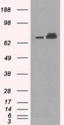
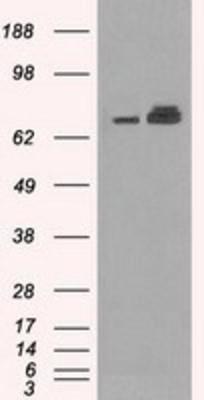
- Western Blot: GRP75/HSPA9B/Mortalin Antibody (9F8) [NBP1-47801] - HEK293T cells were transfected with the pCMV6-ENTRY control (Left lane) or pCMV6-ENTRY GRP75/HSPA9B/Mortalin (Right lane) cDNA for 48 hrs and lysed. Equivalent amounts of cell lysates (5 ug per lane) were separated by SDS-PAGE and immunoblotted with anti-GRP75/HSPA9B/Mortalin.
- Submitted by
- Novus Biologicals (provider)
- Main image

- Experimental details
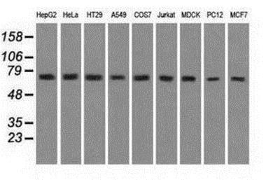
- Western Blot: GRP75/HSPA9B/Mortalin Antibody (9F8) [NBP1-47801] - Analysis of extracts from 9 different cell lines: (HepG2: human; HeLa: human; SVT2: mouse; A549: human; COS7: monkey; Jurkat: human; MDCK: canine; PC12: rat; MCF7: human).
Supportive validation
- Submitted by
- Novus Biologicals (provider)
- Main image

- Experimental details
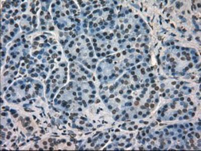
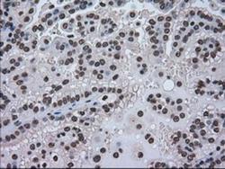
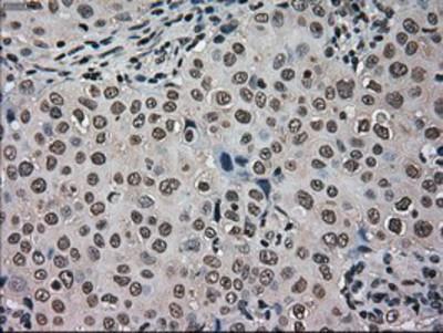
- Immunohistochemistry-Paraffin: GRP75/HSPA9B/Mortalin Antibody (9F8) [NBP1-47801] - Staining of paraffin-embedded Human pancreas tissue using anti-GRP75/HSPA9B/Mortalin mouse monoclonal antibody.
- Submitted by
- Novus Biologicals (provider)
- Main image
- Experimental details
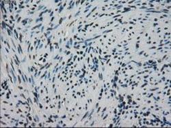
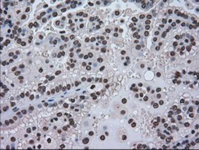
- Immunohistochemistry-Paraffin: GRP75/HSPA9B/Mortalin Antibody (9F8) [NBP1-47801] - Staining of paraffin-embedded Human endometrium tissue using anti-GRP75/HSPA9B/Mortalin mouse monoclonal antibody.

- Submitted by
- Novus Biologicals (provider)
- Main image
- Experimental details
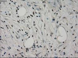
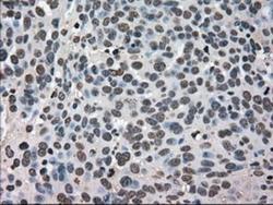
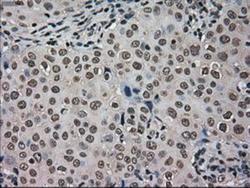
- Immunohistochemistry-Paraffin: GRP75/HSPA9B/Mortalin Antibody (9F8) [NBP1-47801] - Staining of paraffin-embedded Carcinoma of Human pancreas tissue using anti-GRP75/HSPA9B/Mortalin mouse monoclonal antibody.
- Submitted by
- Novus Biologicals (provider)
- Main image
- Experimental details
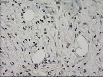
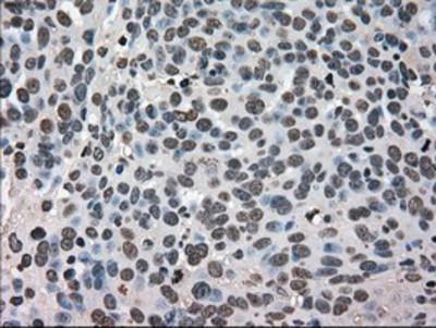
- Immunohistochemistry-Paraffin: GRP75/HSPA9B/Mortalin Antibody (9F8) [NBP1-47801] - Staining of paraffin-embedded Carcinoma of Human kidney tissue using anti-GRP75/HSPA9B/Mortalin mouse monoclonal antibody.
- Submitted by
- Novus Biologicals (provider)
- Main image
- Experimental details
- Immunohistochemistry-Paraffin: GRP75/HSPA9B/Mortalin Antibody (9F8) [NBP1-47801] - Staining of paraffin-embedded Adenocarcinoma of Human ovary tissue using anti-GRP75/HSPA9B/Mortalin mouse monoclonal antibody.
- Submitted by
- Novus Biologicals (provider)
- Main image
- Experimental details
- Immunohistochemistry-Paraffin: GRP75/HSPA9B/Mortalin Antibody (9F8) [NBP1-47801] - Staining of paraffin-embedded Adenocarcinoma of Human breast tissue using anti-GRP75/HSPA9B/Mortalin mouse monoclonal antibody.
Supportive validation
- Submitted by
- Novus Biologicals (provider)
- Main image

- Experimental details
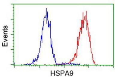
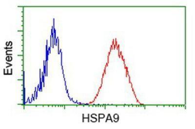
- Flow Cytometry: GRP75/HSPA9B/Mortalin Antibody (9F8) [NBP1-47801] - Analysis of Jurkat cells, using anti-Mortalin antibody, (Red), compared to a nonspecific negative control antibody (Blue).
- Submitted by
- Novus Biologicals (provider)
- Main image

- Experimental details
- Flow Cytometry: GRP75/HSPA9B/Mortalin Antibody (9F8) [NBP1-47801] - Analysis of Hela cells, using anti-Mortalin antibody, (Red), compared to a nonspecific negative control antibody (Blue).