Explore
 Validate
 Learn
Learn Western blot
Antibody data
- Antibody Data
- Antigen structure
- References [0]
- Comments [0]
- Validations
- Western blot [1]
- Immunocytochemistry [1]
- Immunohistochemistry [2]
Submit
Validation data
Reference
Comment
Report error
- Product number
- AF3584 - Provider product page

- Provider
- R&D Systems
- Product name
- Human/Mouse/Rat GRP75/HSPA9B Antibody
- Antibody type
- Polyclonal
- Description
- Immunogen affinity purified. Detects human, mouse, and rat GRP75.
- Reactivity
- Human, Mouse, Rat
- Host
- Goat
- Conjugate
- Unconjugated
- Antigen sequence
P38646- Isotype
- IgG
- Vial size
- 100 ug
- Concentration
- LYOPH
- Storage
- Use a manual defrost freezer and avoid repeated freeze-thaw cycles. 12 months from date of receipt, -20 to -70 °C as supplied. 1 month, 2 to 8 °C under sterile conditions after reconstitution. 6 months, -20 to -70 °C under sterile conditions after reconstitution.
No comments: Submit comment
Supportive validation
- Submitted by
- R&D Systems (provider)
- Main image
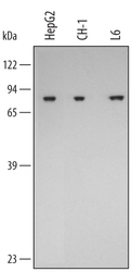
- Experimental details
- Detection of Human/Mouse/Rat GRP75/HSPA9B by Western Blot. Western blot shows lysates of HepG2 human hepatocellular carcinoma cell line, CH-1 mouse B cell lymphoma cell line, and L6 rat myoblast cell line. PVDF membrane was probed with 0.5 µg/mL of Goat Anti-Human/Mouse/Rat GRP75/HSPA9B Antigen Affinity-purified Polyclonal Antibody (Catalog # AF3584) followed by HRP-conjugated Anti-Goat IgG Secondary Antibody (Catalog # HAF109). A specific band was detected for GRP75/HSPA9B at approximately 75 kDa (as indicated). This experiment was conducted under reducing conditions and using Immunoblot Buffer Group 2.

Supportive validation
- Submitted by
- R&D Systems (provider)
- Main image
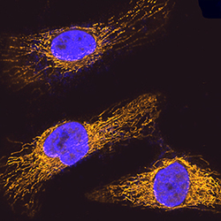
- Experimental details
- GRP75/HSPA9B in HeLa Human Cell Line. GRP75/HSPA9B was detected in immersion fixed HeLa human cervical epithelial carcinoma cell line using Goat Anti-Human/Mouse/Rat GRP75/HSPA9B Antigen Affinity-purified Polyclonal Antibody (Catalog # AF3584) at 5 µg/mL for 3 hours at room temperature. Cells were stained using the NorthernLights™ 557-conjugated Anti-Goat IgG Secondary Antibody (yellow; Catalog # NL001) and counterstained with DAPI (blue). Specific staining was localized to mitochondria. View our protocol for Fluorescent ICC Staining of Cells on Coverslips.

Supportive validation
- Submitted by
- R&D Systems (provider)
- Main image
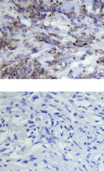
- Experimental details
- GRP75/HSPA9B in Human Meningioma. GRP75/HSPA9B was detected in immersion fixed paraffin-embedded sections of human meningioma using 15 µg/mL Goat Anti-Human/Mouse/Rat GRP75/HSPA9B Antigen Affinity-purified Polyclonal Antibody (Catalog # AF3584) overnight at 4 °C. Tissue was stained with the Anti-Goat HRP-DAB Cell & Tissue Staining Kit (brown; Catalog # CTS008) and counterstained with hematoxylin (blue). Lower panel shows a lack of labeling if primary antibodies are omitted and tissue is stained only with secondary antibody followed by incubation with detection reagents. View our protocol for Chromogenic IHC Staining of Paraffin-embedded Tissue Sections.

- Submitted by
- R&D Systems (provider)
- Main image
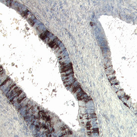
- Experimental details
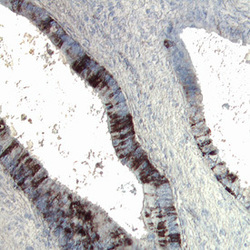
- GRP75/HSPA9B in Human Colon Cancer Tissue. GRP75/HSPA9B was detected in immersion fixed paraffin-embedded sections of human colon cancer tissue using Goat Anti-Human/Mouse/Rat GRP75/HSPA9B Antigen Affinity-purified Polyclonal Antibody (Catalog # AF3584) at 15 µg/mL overnight at 4 °C. Tissue was stained using the Anti-Goat HRP-DAB Cell & Tissue Staining Kit (brown; Catalog # CTS008) and counterstained with hematoxylin (blue). Specific labeling was localized to the plasma membrane of epithelial cells. View our protocol for Chromogenic IHC Staining of Paraffin-embedded Tissue Sections.