Explore
 Validate
 Learn
Learn Western blot
 ELISA
ELISAAntibody data
- Antibody Data
- Antigen structure
- References [3]
- Comments [0]
- Validations
- Western blot [2]
- Immunohistochemistry [1]
Submit
Validation data
Reference
Comment
Report error
- Product number
- GTX26050 - Provider product page

- Provider
- GeneTex
- Proper citation
- GeneTex Cat#GTX26050, RRID:AB_425213
- Product name
- GLI3 antibody
- Antibody type
- Polyclonal
- Reactivity
- Human, Canine, Chicken/Avian, Simian, Xenopus, Zebrafish
- Host
- Rabbit
Submitted references Non-canonical activation of hedgehog in prostate cancer cells mediated by the interaction of transcriptionally active androgen receptor proteins with Gli3.
Generation and characterization of mouse monoclonal antibody 5E1 against human transcription factor GLI3.
Generation and characterization of mouse monoclonal antibody 5E1 against human transcription factor GLI3.
Li N, Truong S, Nouri M, Moore J, Al Nakouzi N, Lubik AA, Buttyan R
Oncogene 2018 Apr;37(17):2313-2325
Oncogene 2018 Apr;37(17):2313-2325
Generation and characterization of mouse monoclonal antibody 5E1 against human transcription factor GLI3.
Hunt R, Bragina O, Drews M, Kasak L, Timmusk S, Valkna A, Kogerman P, Järvekülg L
Hybridoma (2005) 2007 Jun;26(3):131-9
Hybridoma (2005) 2007 Jun;26(3):131-9
Generation and characterization of mouse monoclonal antibody 5E1 against human transcription factor GLI3.
Hunt R, Bragina O, Drews M, Kasak L, Timmusk S, Valkna A, Kogerman P, Järvekülg L
Hybridoma (2005) 2007 Aug;26(4):231-40
Hybridoma (2005) 2007 Aug;26(4):231-40
No comments: Submit comment
Supportive validation
- Submitted by
- GeneTex (provider)
- Main image
- Experimental details
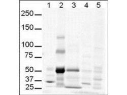
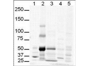
- Western Blot of Rabbit anti-Gli-3 antibody (GTX26050). Lane 1: human brain whole cell lysate. Lane 2: human lung whole cell lysate. Lane 3: human spleen whole cell lysate. Lane 4: mouse brain whole cell lysate. Lane 5: mouse lung whole cell lysate. Load: 20 µg per lane. Primary antibody: Gli-3 antibody at 1:500 for overnight at 4°C. Secondary antibody: infrared rabbit secondary antibody at 1:10,000 for 45 min at RT. Block: 5% BLOTTO overnight at 4°C. Predicted/Observed size: Isoforms at ~170-190kDa and ~80kDa. Lane 2 shows what may be truncated Gli-3 (~80kDa). Other band(s): The strong band at ~50 kDa is unknown.
- Validation comment
- WB
- Submitted by
- GeneTex (provider)
- Main image

- Experimental details
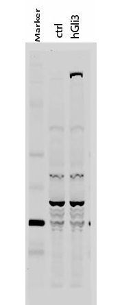
- Western Blot of Rabbit anti-Gli-3 antibody. Lane 1: 50 kDa molecular weight marker. Lane 2: 293T cells transfected with CrkL-Flag. Lane 3: 293T cells transfected with human Gli-3. Load: 35 ?g per lane. Primary antibody: Gli-3 antibody at 1:400 for overnight at 4¢XC. Secondary antibody: IRDye800? rabbit secondary antibody at 1:10,000 for 45 min at RT. Block: 5% BLOTTO overnight at 4¢XC. Predicted/Observed size: 170-190 kDa for hGli-3. Other band(s): Non specific background ~60kDa.
Supportive validation
- Submitted by
- GeneTex (provider)
- Main image

- Experimental details
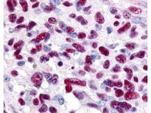
- Immunohistochemistry of Rabbit anti-Gli-3 antibody (GTX26050). This image tissue: human glioblastoma. Specific staining was also noted in tissue from adrenal, brain, glioblastoma, colon, heart, kidney, lung, liver, skeletal muscle, ovary, pancreas, placenta, skin, spleen, stomach, testes, thymus, thyroid, tonsil and uterus. Fixation: formalin fixed paraffin embedded. Antigen retrieval: not required. Primary antibody: Gli-3 antibody at 0.625 ?g/ml for 1 h at RT. Secondary antibody: Peroxidase rabbit secondary antibody at 1:10,000 for 45 min at RT. Localization: Gli-3 is nuclear and smooth muscle. Staining: Gli-3 as precipitated red signal with hematoxylin purple nuclear counterstain.