 Explore
 Validate
 Learn
Learn600-401-694
antibody from Invitrogen Antibodies
Targeting: GLI3
ACLS, GCPS, PAP-A, PAPA, PAPA1, PAPB, PHS, PPDIV
 Western blot
 ELISA
ELISAAntibody data
- Antibody Data
- Antigen structure
- References [0]
- Comments [0]
- Validations
- Western blot [2]
- Immunocytochemistry [2]
- Immunohistochemistry [1]
Submit
Validation data
Reference
Comment
Report error
- Product number
- 600-401-694 - Provider product page

- Provider
- Invitrogen Antibodies
- Product name
- GLI3 Polyclonal Antibody
- Antibody type
- Polyclonal
- Antigen
- Synthetic peptide
- Reactivity
- Human, Canine, Chicken/Avian, Xenopus
- Host
- Rabbit
- Isotype
- IgG
- Vial size
- 100 µg
- Concentration
- 1 mg/mL
- Storage
- -20° C, Avoid Freeze/Thaw Cycles
No comments: Submit comment
Supportive validation
- Submitted by
- Invitrogen Antibodies (provider)
- Main image
- Experimental details
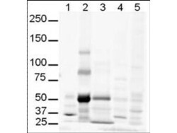
- Western Blot of Rabbit anti-Gli-3 antibody. Lane 1: human brain whole cell lysate. Lane 2: human lung whole cell lysate. Lane 3: human spleen whole cell lysate. Lane 4: mouse brain whole cell lysate. Lane 5: mouse lung whole cell lysate. Load: 20 µg per lane. Primary antibody: Gli-3 antibody at 1:500 for overnight at 4°C. Secondary antibody: IRDye800™ rabbit secondary antibody at 1:10,000 for 45 min at RT. Block: 5% BLOTTO overnight at 4°C. Predicted/Observed size: Isoforms at ~170-190kDa and ~80kDa. Lane 2 shows what may be truncated Gli-3 (~80kDa). Other band(s): The strong band at ~50 kDa is unknown.

- Submitted by
- Invitrogen Antibodies (provider)
- Main image

- Experimental details
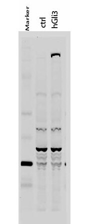
- Western Blot of Rabbit anti-Gli-3 antibody. Lane 1: 50 kDa molecular weight marker. Lane 2: 293T cells transfected with CrkL-Flag. Lane 3: 293T cells transfected with human Gli-3. Load: 35 µg per lane. Primary antibody: Gli-3 antibody at 1:400 for overnight at 4°C. Secondary antibody: IRDye800™ rabbit secondary antibody at 1:10,000 for 45 min at RT. Block: 5% BLOTTO overnight at 4°C. Predicted/Observed size: 170-190 kDa for hGli-3. Other band(s): Non specific background ~60kDa.
Supportive validation
- Submitted by
- Invitrogen Antibodies (provider)
- Main image

- Experimental details
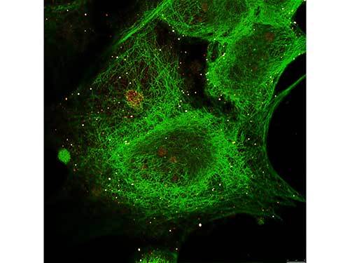
- Immunofluorescence Microscopy of Rabbit anti-Gli-3 antibody. Tissue: MCF-7 cell. Antigen retrieval: not required. Primary antibody: Gli-3 antibody and Anti-alpha-Tubulin at 5 µg/mL for 1 h at RT. Secondary antibody: Fluorescein secondary antibody at 1:10,000 for 45 min at RT. Localization: Gli-3 is nuclear. Staining: Gli-3 staining as red fluorescent signal and Anti-alpha-Tubulin staining as green fluorescent signal using STED.
- Submitted by
- Invitrogen Antibodies (provider)
- Main image
- Experimental details
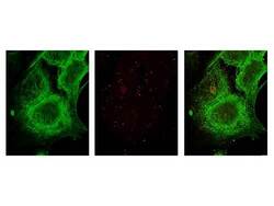
- Immunofluorescence Microscopy of Rabbit anti-Gli-3 antibody. Tissue: MCF-7 cell. Antigen retrieval: not required. Primary antibody: Gli-3 antibody and Anti-alpha-Tubulin at 5 µg/mL for 1 h at RT. Secondary antibody: Fluorescein secondary antibody at 1:10,000 for 45 min at RT. Localization: Gli-3 is nuclear. Staining: Image (1) shows alpha-Tubulin staining as green fluorescent signal. Image (2) shows Gli-3 staining as red fluorescent signal and Images (3) shows both antibodies fluorescing using STED microscopy.

Supportive validation
- Submitted by
- Invitrogen Antibodies (provider)
- Main image
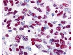
- Experimental details
- Immunohistochemistry of Rabbit anti-Gli-3 antibody. This image tissue: human glioblastoma. Specific staining was also noted in tissue from adrenal, brain, glioblastoma, colon, heart, kidney, lung, liver, skeletal muscle, ovary, pancreas, placenta, skin, spleen, stomach, testes, thymus, thyroid, tonsil and uterus. Fixation: formalin fixed paraffin embedded. Antigen retrieval: not required. Primary antibody: Gli-3 antibody at 0.625 µg/ml for 1 h at RT. Secondary antibody: Peroxidase rabbit secondary antibody at 1:10,000 for 45 min at RT. Localization: Gli-3 is nuclear and smooth muscle. Staining: Gli-3 as precipitated red signal with hematoxylin purple nuclear counterstain.
