Explore
 Validate
 Learn
Learn Western blot
Antibody data
- Antibody Data
- Antigen structure
- References [0]
- Comments [0]
- Validations
- Western blot [4]
- Immunocytochemistry [2]
- Immunohistochemistry [3]
Submit
Validation data
Reference
Comment
Report error
- Product number
- PA5-27949 - Provider product page

- Provider
- Invitrogen Antibodies
- Product name
- Cytokeratin 17 Polyclonal Antibody
- Antibody type
- Polyclonal
- Antigen
- Recombinant protein fragment
- Description
- Recommended positive controls: HeLa, Rat bladder. Predicted reactivity: Mouse (96%), Rat (96%), Sheep (95%), Chimpanzee (99%), Bovine (95%). Store product as a concentrated solution. Centrifuge briefly prior to opening the vial.
- Reactivity
- Human, Mouse, Rat
- Host
- Rabbit
- Isotype
- IgG
- Vial size
- 100 µL
- Concentration
- 1 mg/mL
- Storage
- Store at 4°C short term. For long term storage, store at -20°C, avoiding freeze/thaw cycles.
No comments: Submit comment
Supportive validation
- Submitted by
- Invitrogen Antibodies (provider)
- Main image
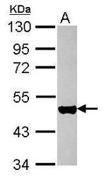
- Experimental details
- Western blot analysis of Cytokeratin 17 using 30 µg of HeLa lysate. Samples were loaded onto a 10% SDS-PAGE gel and probed with a Cytokeratin 17 polyclonal antibody (Product # PA5-27949) at a dilution of 1:10,000.

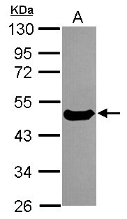
- Submitted by
- Invitrogen Antibodies (provider)
- Main image

- Experimental details
- Western Blot using Cytokeratin 17 Polyclonal Antibody (Product # PA5-27949). Sample (50 µg of whole cell lysate). Lane A: Rat bladder. 10% SDS PAGE. Cytokeratin 17 Polyclonal Antibody (Product # PA5-27949) diluted at 1:10,000.
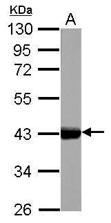
- Submitted by
- Invitrogen Antibodies (provider)
- Main image

- Experimental details
- Western Blot using Cytokeratin 17 Polyclonal Antibody (Product # PA5-27949). Sample (30 µg of whole cell lysate). Lane A: HeLa. 10% SDS PAGE. Cytokeratin 17 Polyclonal Antibody (Product # PA5-27949) diluted at 1:5,000.
- Submitted by
- Invitrogen Antibodies (provider)
- Main image
- Experimental details
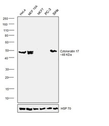
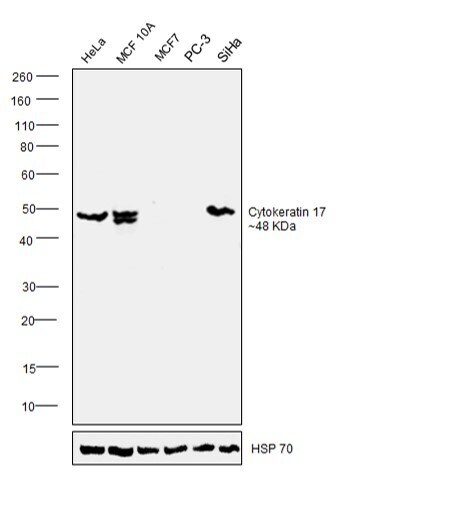
- Western blot was performed using Anti- Cytokeratin 17 Polyclonal Antibody (Product # PA5-27949) and a 48 kDa band corresponding to Cytokeratin 17 was observed across cell lines tested except MCF7 and PC-3. Whole cell extracts (30 µg lysate) of HeLa (Lane 1), MCF 10A (Lane 2), MCF-7 (Lane 3), PC-3 (Lane 4) and SiHa (Lane 5) were electrophoresed using NuPAGE™ 4-12% Bis-Tris Protein Gel (Product # NP0322BOX). Resolved proteins were then transferred onto a nitrocellulose membrane (Product # IB23001) by iBlot® 2 Dry Blotting System (Product # IB21001). The blot was probed with the primary antibody (1:10000) and detected by chemiluminescence with Goat anti-Rabbit IgG (H+L) Superclonal™ Recombinant Secondary Antibody, HRP (Product # A27036, 1:4000 dilution) using the iBright FL 1000 (Product # A32752). Chemiluminescent detection was performed using Novex® ECL Chemiluminescent Substrate Reagent Kit (Product # WP20005).
Supportive validation
- Submitted by
- Invitrogen Antibodies (provider)
- Main image
- Experimental details
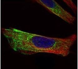
- Immunofluorescent analysis of Cytokeratin 17 in paraformaldehyde-fixed HeLa cells using a Cytokeratin 17 polyclonal antibody (Product # PA5-27949) (Green) at a 1:500 dilution. Alpha-tubulin filaments were labeled with Product # PA5-29281 (Red) at a 1:2500.

- Submitted by
- Invitrogen Antibodies (provider)
- Main image

- Experimental details
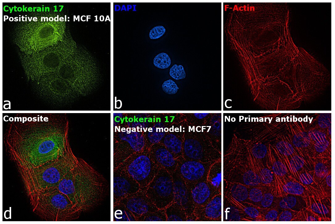
- Immunofluorescence analysis of Cytokeratin 17 was performed using MCF 10A and MCF7 cells. The cells were fixed with 4% paraformaldehyde for 10 minutes, permeabilized with 0.1% Triton™ X-100 for 15 minutes, and blocked with 2% BSA for 1 hour at room temperature. The cells were labeled with Cytokeratin 17 Polyclonal Antibody (Product # PA5-27949) at 1:100 dilution in 0.1% BSA and incubated overnight at 4 degree and then labeled with Donkey anti-Rabbit IgG (H+L) Highly Cross-Adsorbed Secondary Antibody, Alexa Fluor Plus 488 conjugate (Product # A32790) at a dilution of 1:2000 for 45 minutes at room temperature (Panel a: green). Nuclei (Panel b: blue) were stained with ProLong™ Diamond Antifade Mountant with DAPI (Product # P36962). F-actin (Panel c: red) was stained with Rhodamine Phalloidin (Product # R415, 1:300). Panel d represents the composite image showing cytoskeletal localization of Keratin 17 in MCF 10A cells but not in MCF7 cells (panel e) which are reported to be low to negative for its expression. Panel f represents control cells with no primary antibody to assess background. The images were captured at 60X magnification.
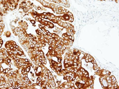
Supportive validation
- Submitted by
- Invitrogen Antibodies (provider)
- Main image
- Experimental details
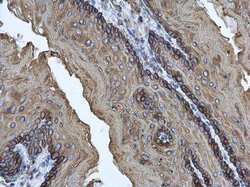
- Immunohistochemistry (Paraffin) analysis of Cytokeratin 17 was performed in paraffin-embedded mouse esophagus tissue using Cytokeratin 17 Polyclonal Antibody (Product # PA5-27949) at a dilution of 1:500.

- Submitted by
- Invitrogen Antibodies (provider)
- Main image
- Experimental details
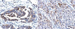
- Immunohistochemistry (Paraffin) analysis of Cytokeratin 17 was performed in paraffin-embedded human cervix tissue using Cytokeratin 17 Polyclonal Antibody (Product # PA5-27949) at a dilution of 1:500.

- Submitted by
- Invitrogen Antibodies (provider)
- Main image
- Experimental details
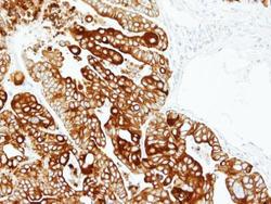
- Immunohistochemical analysis of paraffin-embedded N87 xenograft, using Cytokeratin 17 (Product # PA5-27949) antibody at 1:100 dilution. Antigen Retrieval: EDTA based buffer, pH 8.0, 15 min.