Explore
 Validate
 Learn
Learn Western blot
 Immunoprecipitation
Antibody data
- Antibody Data
- Antigen structure
- References [0]
- Comments [0]
- Validations
- Western blot [1]
- Immunohistochemistry [4]
- Other assay [1]
Submit
Validation data
Reference
Comment
Report error
- Product number
- MA1-19045 - Provider product page

- Provider
- Invitrogen Antibodies
- Product name
- Cytokeratin 7/17 Monoclonal Antibody (C-46)
- Antibody type
- Monoclonal
- Antigen
- Other
- Description
- This antibody reacts with cytokeratin peptides 7 and 17 (54 and 46 kDa). Cytokeratins are members of intermediate filaments subfamily of intracellular proteins represented in epithelial tissues. This antibody will not cross-react with mouse, rat or rabbit.
- Reactivity
- Human, Bovine, Porcine
- Host
- Mouse
- Isotype
- IgG
- Antibody clone number
- C-46
- Vial size
- 100 µg
- Concentration
- 1 mg/mL
- Storage
- 4° C, do not freeze
No comments: Submit comment
Supportive validation
- Submitted by
- Invitrogen Antibodies (provider)
- Main image

- Experimental details
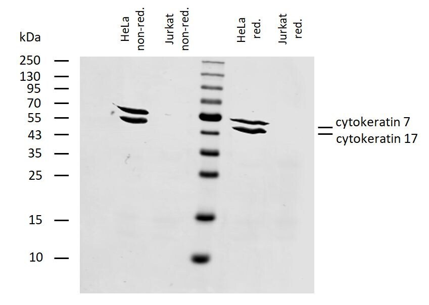
- Western blotting analysis of human cytokeratin 7+17 using mouse Monoclonal antibody C-46 (Product # MA1-19045) on lysates of HeLa cell line and Jurkat cell line (cytokeratin non-expressing cell line; negative control) under non-reducing and reducing conditions. Nitrocellulose membrane was probed with 2µg/mL of mouse anti-cytokeratin 7+17 monoclonal antibody C-46 followed by IRDye800-conjugated anti-mouse IgG1 secondary antibody. A specific band was detected for cytokeratin 17 at approximately 46kDa and for cytokeratin 7 at approximately 54 kDa.
Supportive validation
- Submitted by
- Invitrogen Antibodies (provider)
- Main image
- Experimental details
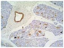
- Immunohistochemical analysis of Cytokeratin 7/17 using a monoclonal antibody (Product # MA1-19045).

- Submitted by
- Invitrogen Antibodies (provider)
- Main image
- Experimental details
- Immunohistochemical analysis of Cytokeratin 7/17 using a monoclonal antibody (Product # MA1-19045).
- Submitted by
- Invitrogen Antibodies (provider)
- Main image

- Experimental details
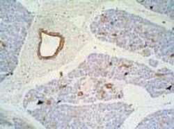
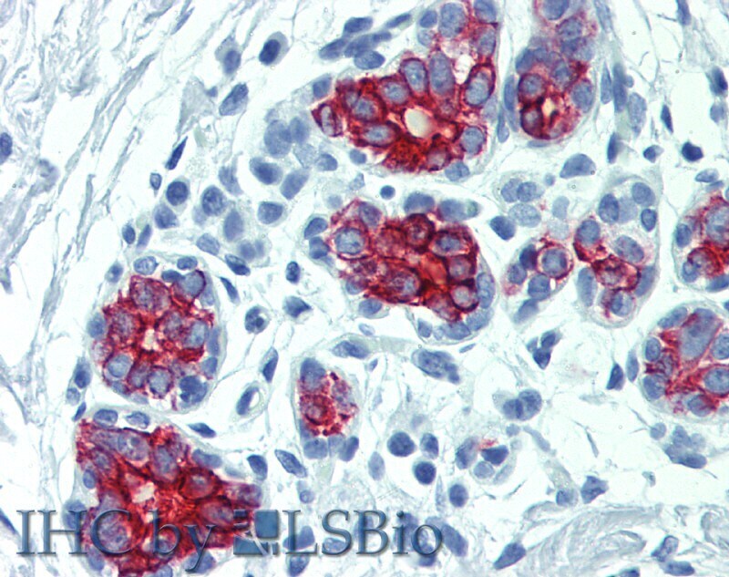
- Immunohistochemistry staining of human breast (paraffin-embedded sections) with anti-cytokeratin 7+17 (C-46) Monoclonal antibody (Product # MA1-19045).
- Submitted by
- Invitrogen Antibodies (provider)
- Main image

- Experimental details
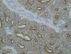
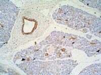
- Immunohistochemistry staining of pancreas (paraffin-embedded sections) with anti-Cytokeratin 7+17 (C-46) Monoclonal antibody (Product # MA1-19045).
Supportive validation
- Submitted by
- Invitrogen Antibodies (provider)
- Main image
- Experimental details
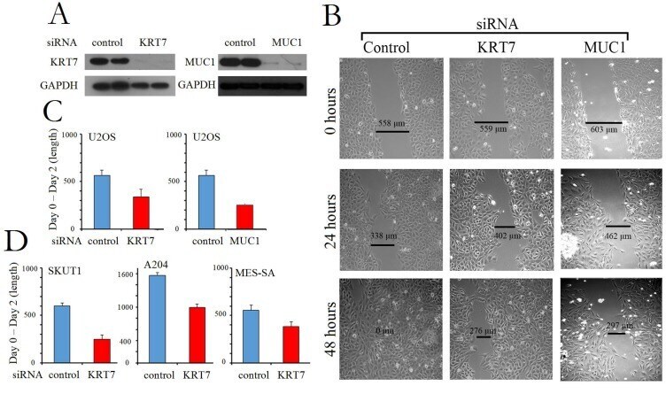
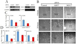
- Figure 4 Gene knockdown of KRT7 and MUC1 blocks metastasis. ( A ) Western blot of U2OS sarcoma cell line subjected to gene knockdown using siRNA directed at KRT7 (Left panel) and MUC1 (Right panel). GAPDH was used as a loading control. ( B ) Cell migration/wound healing assay on U2OS osteosarcoma cells from ( A ) were analyzed at 0, 24, and 48 hrs (Left panel: control siRNA; Center panel: KRT7 siRNA; Right panel: MUC1 siRNA). Representative images from three independent experiments in osteosarcoma U2OS cells are presented and quantified in ( C ). ( D ) Relative cell migration/wound healing in several cell lines: leiomyosarcoma SKUT1 cells, rhabdoid tumor A204 cells, and uterine sarcoma MES-SA cells. The length at Day 2 is subtracted from the length at Day 0. The error bars represent standard deviation. Representative calculations from three independent experiments are presented.