 Explore
 Validate
 Learn
Learn Western blot
 Immunocytochemistry
 Immunoprecipitation
Antibody data
- Antibody Data
- Antigen structure
- References [0]
- Comments [0]
- Validations
- Western blot [2]
- Immunoprecipitation [3]
- Immunohistochemistry [2]
Submit
Validation data
Reference
Comment
Report error
- Product number
- LS-C96661 - Provider product page

- Provider
- LSBio
- Proper citation
- LifeSpan Cat#LS-C96661, RRID:AB_2117079
- Product name
- HDAC9 Antibody (aa2-32) LS-C96661
- Antibody type
- Polyclonal
- Description
- Ammonium sulfate precipitation
- Reactivity
- Human
- Host
- Rabbit
- Storage
- Maintain refrigerated at 2°C to 8°C for up to 6 months. For long term storage store at -20°C.
No comments: Submit comment
Enhanced validation
- Submitted by
- LSBio (provider)
- Enhanced method
- Genetic validation
- Main image

- Experimental details
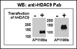
- Both anti-HDAC9 N-term and C-term antibody were tested by WB and IP-WB using HeLa and HeLa-HDAC9 transfected cells. Top figure shows both antibody specifically detect HDAC9 in HeLa-HDAC9 transfected cell but not HeLa alone.
- Submitted by
- LSBio (provider)
- Enhanced method
- Genetic validation
- Main image
- Experimental details
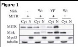
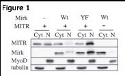
- Figure 1: Immunoblots for MITR / HDAC9 N-term antibody), Mirk, MyoD and tubulin proteins are shown for cytoplasmic (Cyt) and nuclear (N) extracts from undifferentiated C2C12 myoblasts. Before cell collection for fractionation, the cells are transfected with plasmids coding for Mirk (Wt), kinase-inactive Mirk (YF) or MITR. Data courtesy of laboratory of Dr. Eileen Friedman. Dept of Pathology, Upstate Medical University, State University of New York.
Supportive validation
- Submitted by
- LSBio (provider)
- Enhanced method
- Genetic validation
- Main image

- Experimental details
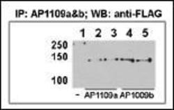
- This figure shows that both antibody can immunoprecipitate (IP) HDAC9 from HeLa-HDAC9 transfected cells. (Data kindly provided by Dr. Zhigang Yuan, H. Lee Moffitt Cancer Center and Research Institute, Tampa, FL).

- Submitted by
- LSBio (provider)
- Main image
- Experimental details
- This figure shows that both antibody can immunoprecipitate (IP) HDAC9 from HeLa-HDAC9 transfected cells. (Data kindly provided by Dr. Zhigang Yuan, H. Lee Moffitt Cancer Center and Research Institute, Tampa, FL).

- Submitted by
- LSBio (provider)
- Main image

- Experimental details
- This figure shows that both antibody can immunoprecipitate (IP) HDAC9 from HeLa-HDAC9 transfected cells. (Data kindly provided by Dr. Zhigang Yuan, H. Lee Moffitt Cancer Center and Research Institute, Tampa, FL).

Enhanced validation
- Submitted by
- LSBio (provider)
- Enhanced method
- Genetic validation
- Main image
- Experimental details
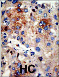
- Formalin-fixed and paraffin-embedded human cancer tissue reacted with the primary antibody, which was peroxidase-conjugated to the secondary antibody, followed by AEC staining. This data demonstrates the use of this antibody for immunohistochemistry; clinical relevance has not been evaluated. BC = breast carcinoma; HC = hepatocarcinoma.

- Submitted by
- LSBio (provider)
- Main image

- Experimental details
- Formalin-fixed and paraffin-embedded human cancer tissue reacted with the primary antibody, which was peroxidase-conjugated to the secondary antibody, followed by AEC staining. This data demonstrates the use of this antibody for immunohistochemistry; clinical relevance has not been evaluated. BC = breast carcinoma; HC = hepatocarcinoma.
