Explore
 Validate
 Learn
Learn Western blot
Antibody data
- Antibody Data
- Antigen structure
- References [3]
- Comments [0]
- Validations
- Western blot [1]
- Immunocytochemistry [1]
- Immunoprecipitation [1]
- Immunohistochemistry [1]
Submit
Validation data
Reference
Comment
Report error
- Product number
- PAB2338 - Provider product page

- Provider
- Abnova Corporation
- Proper citation
- Abnova Corporation Cat#PAB2338, RRID:AB_1575875
- Product name
- HDAC9 polyclonal antibody
- Antibody type
- Polyclonal
- Description
- Rabbit polyclonal antibody raised against synthetic peptide of HDAC9.
- Storage
- Store at 4°C. For long term storage store at -20°C.Aliquot to avoid repeated freezing and thawing.
Submitted references Molecular characterization of a familial translocation implicates disruption of HDAC9 and possible position effect on TGFbeta2 in the pathogenesis of Peters' anomaly.
The histone deacetylase 9 gene encodes multiple protein isoforms.
Chromosomal organization and localization of the human histone deacetylase 9 gene (HDAC9).
David D, Cardoso J, Marques B, Marques R, Silva ED, Santos H, Boavida MG
Genomics 2003 May;81(5):489-503
Genomics 2003 May;81(5):489-503
The histone deacetylase 9 gene encodes multiple protein isoforms.
Petrie K, Guidez F, Howell L, Healy L, Waxman S, Greaves M, Zelent A
The Journal of biological chemistry 2003 May 2;278(18):16059-72
The Journal of biological chemistry 2003 May 2;278(18):16059-72
Chromosomal organization and localization of the human histone deacetylase 9 gene (HDAC9).
Mahlknecht U, Schnittger S, Will J, Cicek N, Hoelzer D
Biochemical and biophysical research communications 2002 Apr 26;293(1):182-91
Biochemical and biophysical research communications 2002 Apr 26;293(1):182-91
No comments: Submit comment
Supportive validation
- Submitted by
- Abnova Corporation (provider)
- Main image
- Experimental details
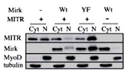
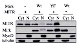
- Immunoblots for HDAC9 polyclonal antibody (Cat # PAB2338), Mirk, MyoD and tubulin proteins are shown for cytoplasmic (Cyt) and nuclear (N) extracts from undifferentiated C2C12 myoblasts. Before cell collection for fractionation, the cells are transfected with plasmids coding for Mirk (Wt), kinase-inactive Mirk (YF) or MITR. Data courtesy of laboratory of Dr. Eileen Friedman. Dept of Pathology, Upstate Medical University, State University of New York.
Supportive validation
- Submitted by
- Abnova Corporation (provider)
- Main image
- Experimental details
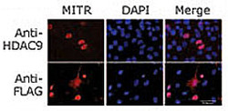
- Immunofluorescence staining of MITR for a compartmentalization study in undifferentiated C2C12 myoblasts transfected with a MITR-expressing plasmid. MITR is detected by using the HDAC9 N-term antibody (top panel) or a FLAG antibody (bottom panel) detecting a FLAG epitope fused at the N-term end of the MITR construct. Data courtesy of laboratory of Dr. Eileen Friedman. Dept of Pathology, Upstate Medical University, State University of New York.
- Validation comment
- Immunofluorescence

Supportive validation
- Submitted by
- Abnova Corporation (provider)
- Main image

- Experimental details
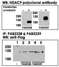
- Both HDAC9 (Cat # PAB2338) and HDAC9 (Cat # PAB2339) polyclonal antibody were tested by WB and IP-WB using HeLa and HeLa-HDAC9 transfected cells. Top figure shows both polyclonal antibody specifically detect HDAC9 in HeLa-HDAC9 transfected cell but not HeLa alone. Bottom figure shows that both polyclonal antibody can immunoprecipitate (IP) HDAC9 from HeLa-HDAC9 tranfected cells. (Data kindly provided by Dr. Zhigang Yuan, H. Lee Moffitt Cancer Center and Research Institute, Tampa, FL) .
- Validation comment
- Immunoprecipitation
Supportive validation
- Submitted by
- Abnova Corporation (provider)
- Main image

- Experimental details
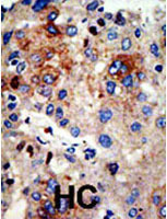
- Formalin-fixed and paraffin-embedded human hepatocellular carcinoma tissue reacted with the primary antibody, which was peroxidase-conjugated to the secondary antibody, followed by AEC staining. This data demonstrates the use of this antibody for immunohistochemistry ; clinical relevance has not been evaluated. HC = hepatocarcinoma.
- Validation comment
- Immunohistochemistry (Formalin/PFA-fixed paraffin-embedded sections)