Explore
 Validate
 Learn
Learn Western blot
 Immunocytochemistry
Antibody data
- Antibody Data
- Antigen structure
- References [0]
- Comments [0]
- Validations
- Immunocytochemistry [3]
- Immunohistochemistry [1]
Submit
Validation data
Reference
Comment
Report error
- Product number
- PA5-112136 - Provider product page

- Provider
- Invitrogen Antibodies
- Product name
- Galectin 7 Polyclonal Antibody
- Antibody type
- Polyclonal
- Antigen
- Recombinant full-length protein
- Reactivity
- Human
- Host
- Rabbit
- Isotype
- IgG
- Vial size
- 100 μL
- Storage
- Store at 4°C short term. For long term storage, store at -20°C, avoiding freeze/thaw cycles.
No comments: Submit comment
Supportive validation
- Submitted by
- Invitrogen Antibodies (provider)
- Main image
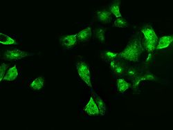
- Experimental details
- Immunocytochemistry-Immunofluorescence analysis of Galectin 7 in A431 cells. Cells were fixed with 4% PFA, permeabilized with 0.3% Triton X-100 in PBS, blocked with 10% serum, and incubated with Galectin 7 Polyclonal Antibody (Product # PA5-112136) (1:5000) at 4°C overnight. Then cells were stained with the Alexa Fluor 488-conjugated Goat Anti-rabbit IgG secondary antibody (green). Positive staining was localized to cytoplasm and nucleus.

- Submitted by
- Invitrogen Antibodies (provider)
- Main image
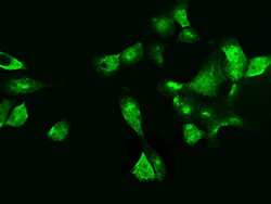
- Experimental details
- Immunocytochemistry-Immunofluorescence analysis of Galectin 7 in A431 cells. Cells were fixed with 4% PFA, permeabilized with 0.3% Triton X-100 in PBS, blocked with 10% serum, and incubated with Galectin 7 Polyclonal Antibody (Product # PA5-112136) (1:5000) at 4°C overnight. Then cells were stained with the Alexa Fluor 488-conjugated Goat Anti-rabbit IgG secondary antibody (green). Positive staining was localized to cytoplasm and nucleus.

- Submitted by
- Invitrogen Antibodies (provider)
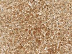
- Main image

- Experimental details
- Immunocytochemistry-Immunofluorescence analysis of Galectin 7 in A431 cells. Cells were fixed with 4% PFA, permeabilized with 0.3% Triton X-100 in PBS, blocked with 10% serum, and incubated with Galectin 7 Polyclonal Antibody (Product # PA5-112136) (1:5000) at 4°C overnight. Then cells were stained with the Alexa Fluor 488-conjugated Goat Anti-rabbit IgG secondary antibody (green). Positive staining was localized to cytoplasm and nucleus.

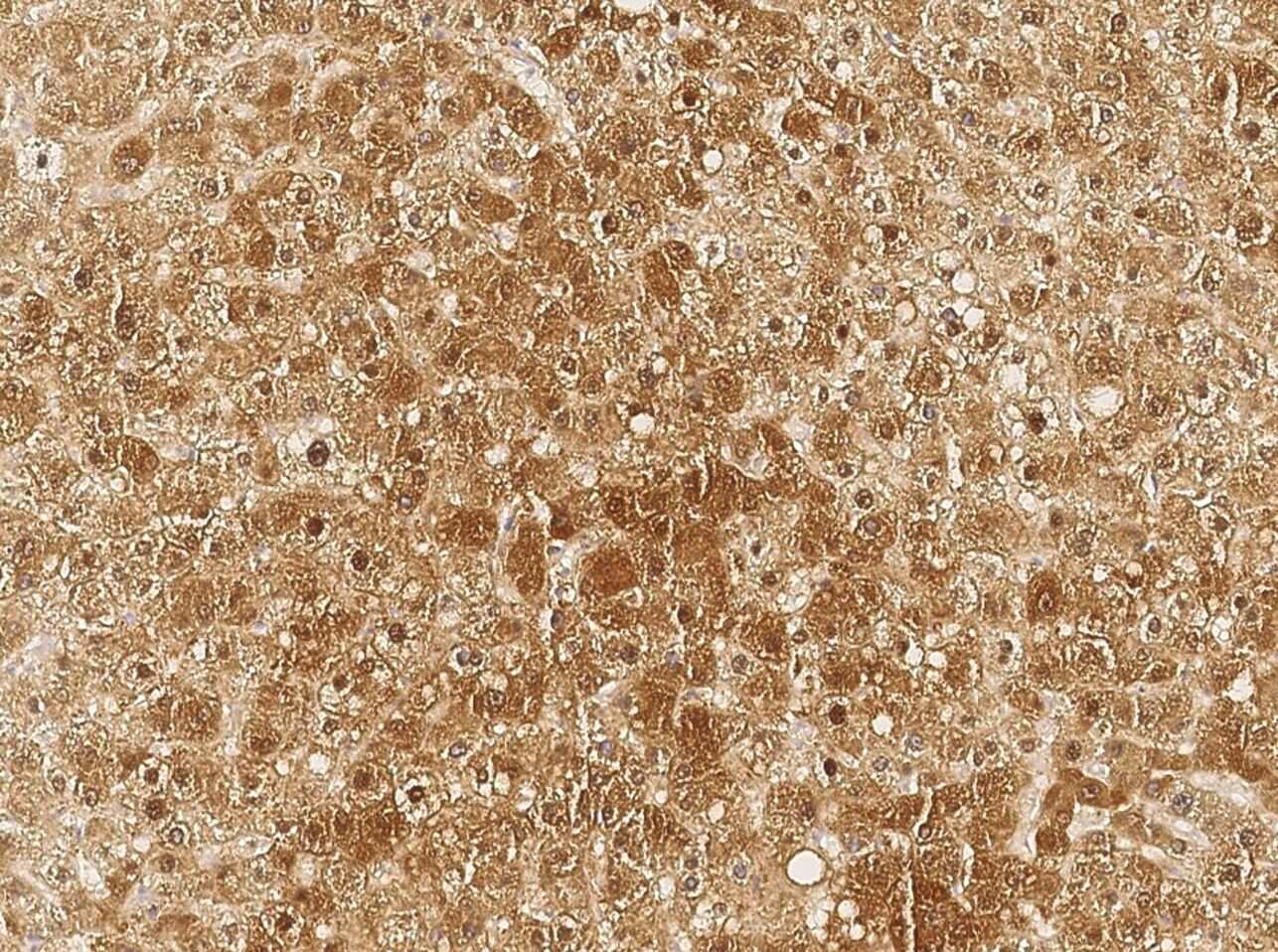
Supportive validation
- Submitted by
- Invitrogen Antibodies (provider)
- Main image
- Experimental details
- Immunohistochemistry analysis of Galectin 7 in formalin-fixed paraffin embedded sections human liver tissue sections using Galectin 7 Polyclonal Antibody (Product # PA5-112136) at a dilution of 1:5000.