Antibody data
- Antibody Data
- Antigen structure
- References [0]
- Comments [0]
- Validations
- Western blot [2]
Submit
Validation data
Reference
Comment
Report error
- Product number
- LS-B329 - Provider product page

- Provider
- LSBio
- Proper citation
- LifeSpan Cat#LS-B329, RRID:AB_2274226
- Product name
- IHC-plus™ ATF3 Antibody LS-B329
- Antibody type
- Polyclonal
- Description
- Affinity purified
- Reactivity
- Human
- Host
- Rabbit
- Isotype
- IgG
- Storage
- Store at 4°C or -20°C. Avoid freeze-thaw cycles.
No comments: Submit comment
Supportive validation
- Submitted by
- LSBio (provider)
- Enhanced method
- Genetic validation
- Main image

- Experimental details
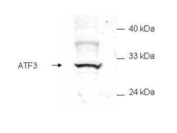
- Anti-ATF3 Antibody - Western Blot. Western blot of mammalian whole cell extract transfected with HA epitope tagged human ATF3. Affinity purified anti-ATF3 detects a band ~31 kD corresponding to recombinant human ATF3. Immunostaining using anti-HA epitope tag antibody confirms the composition of the recombinant band (not shown). The protein was transferred to nitrocellulose in 30 minutes using standard methods. After blocking with 5% goat serum and 0.5% non-fat milk in PBS, the membrane was probed with the primary antibody diluted 1:200 in 0.2X blocking buffer in PBS overnight at 4C. Reaction was followed by washes and reaction with a 1:5000 dilution of IRDye800 conjugated Gt-a-Rabbit IgG [H&L] (code for 30 min at room temperature. LICORs Odyssey Infrared Imaging System was used to scan and process the image. Other detection systems will yield similar results.
- Submitted by
- LSBio (provider)
- Enhanced method
- Genetic validation
- Main image

- Experimental details
- Anti-ATF3 Antibody - Western Blot. Western blot of E. coli whole cell extract transfected with GST epitope tagged human ATF3. Affinity purified anti-ATF3 detects a band ~48 kD corresponding to recombinant human ATF3. Immunostaining using anti-GST epitope tag antibody confirms the composition of the recombinant band (not shown). The protein was transferred to nitrocellulose using standard methods. After blocking with 5% goat serum and 0.5% non fat milk in PBS, the membrane was probed with the primary antibody diluted 1:200 in 0.2X blocking buffer in PBS overnight at 4°C. Reaction was followed by washes and reaction with a 1:5000 dilution of IRDye800 conjugated Gt-a-Rabbit IgG [H&L] (code for 30 min at room temperature. LICORs Odyssey Infrared Imaging System was used to scan and process the image. Other detection systems will yield similar results.
 Explore
 Validate
 Learn
Learn Western blot
 ELISA
ELISA Immunohistochemistry