Explore
 Validate
 Learn
Learn Western blot
 ELISA
ELISAAntibody data
- Antibody Data
- Antigen structure
- References [0]
- Comments [0]
- Validations
- Western blot [2]
Submit
Validation data
Reference
Comment
Report error
- Product number
- AP09241PU-N - Provider product page

- Provider
- Acris Antibodies GmbH
- Proper citation
- Acris Antibodies GmbH Cat#AP09241PU-N, RRID:AB_2035147
- Product name
- anti ATF3 (113-130)
- Antibody type
- Polyclonal
- Antigen
- Other
- Reactivity
- Human
- Host
- Rabbit
- Isotype
- IgG
- Vial size
- 0.1 mg
- Concentration
- 1.0 mg/ml (by UV absorbance at 280 nm)
No comments: Submit comment
Supportive validation
- Submitted by
- Acris Antibodies GmbH (provider)
- Main image
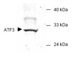
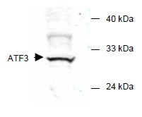
- Experimental details
- Western blot of mammalian whole cell extract transfected with HA epitope tagged human ATF3. Anti-ATF3 detects a band ~31 kDa corresponding to recombinant human ATF3. Immunostaining using anti-HA epitope tag antibody confirms the composition of the recombinant band (not shown). The protein was transferred to nitrocellulose in 30 minutes using standard methods. After blocking with 5% goat serum and 0.5% non-fat milk in PBS, the membrane was probed with the primary antibody diluted 1:200 in 0.2X blocking buffer in PBS overnight at 4°C. Reaction was followed by washes and reaction with a 1:5000 dilution of IRDye(TM)800 conjugated Gt-a-Rabbit IgG [H&L] for 30 min at room temperature. LICOR's Odyssey® Infrared Imaging System was used to scan and process the image. Other detection systems will yield similar results.
- Submitted by
- Acris Antibodies GmbH (provider)
- Main image
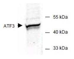
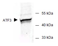
- Experimental details
- Western blot of E.coli whole cell extract transfected with GST epitope tagged human ATF3. Anti-ATF3 detects a band ~48 kDa corresponding to recombinant human ATF3. Anti-GST epitope tag antibodyconfirms the composition of the recombinant band (not shown). The protein was transferred to nitrocellulose using standard methods. Afterblocking with 5% goat serum and 0.5% non fat milk in PBS, the membrane was probed with the primary antibody diluted 1:200 in 0.2X blocking buffer in PBS overnight at 4°C. Reaction was followed bywashes and reaction with a 1:5000 dilution of IRDye(TM)800 conjugated Gta-Rabbit IgG [H&L] for 30 min at room temperature. LICOR's Odyssey® Infrared Imaging System was used to scan and process the image. Other detection systems will yield similar results.