Explore
 Validate
 Learn
Learn Western blot
Antibody data
- Antibody Data
- Antigen structure
- References [1]
- Comments [0]
- Validations
- Western blot [2]
- Other assay [3]
Submit
Validation data
Reference
Comment
Report error
- Product number
- PA5-81166 - Provider product page

- Provider
- Invitrogen Antibodies
- Product name
- E2F5 Polyclonal Antibody
- Antibody type
- Polyclonal
- Antigen
- Synthetic peptide
- Description
- This product is preservative free. It is recommended to add sodium azide to avoid contamination (final concentration 0.05%-0.1%).
- Concentration
- 5 mg/mL
Submitted references An E2F5-TFDP1-BRG1 Complex Mediates Transcriptional Activation of MYCN in Hepatocytes.
Fan Z, Kong M, Miao X, Guo Y, Ren H, Wang J, Wang S, Tang N, Shang L, Zhu Z, Liu H, Zhu W, Shi X
Frontiers in cell and developmental biology 2021;9:742319
Frontiers in cell and developmental biology 2021;9:742319
No comments: Submit comment
Supportive validation
- Submitted by
- Invitrogen Antibodies (provider)
- Main image

- Experimental details
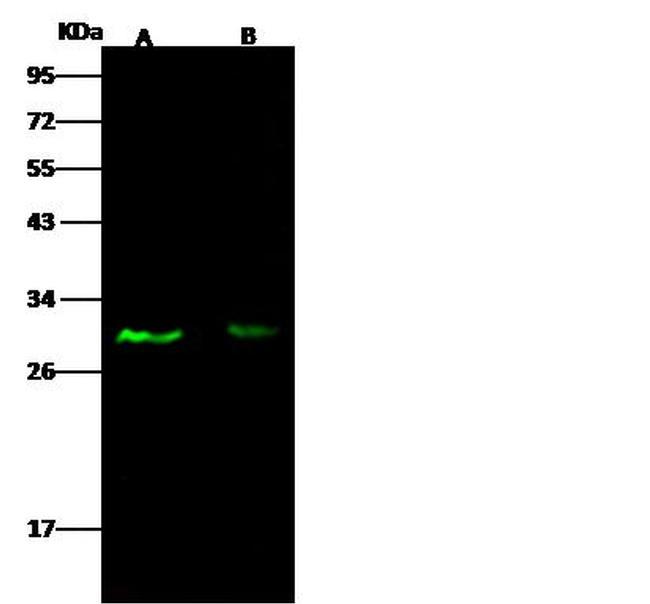
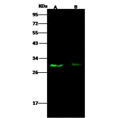
- Western blot analysis of E2F4/E2F5 in Lane A: Jurkat Whole Cell Lysate (30 µg), Lane B: HepG2 Whole Cell Lysate (30 µg) . Samples were probed using a E2F4/E2F5 Polyclonal Antibody (Product # PA5-81166) at a 1:500 dilution, followed by a Goat Anti-Rabbit IgG (H+L), Dylight 800 Secondary Antibody at a 1:10000 dilution. Western blot was performed under reducing conditions. Predicted band size:38 kDa. Observed band size:30 kDa.
- Submitted by
- Invitrogen Antibodies (provider)
- Main image

- Experimental details
- Western Blot using E2F5 Polyclonal Antibody (Product # PA5-81166) at 1:500 dilution. Lane A: Jurkat Whole Cell Lysate, Lane B: HepG2 Whole Cell Lysate. Lysates/proteins at 30 μg per lane. Secondary Goat Anti- RabbitIgG H&L (DyLight™ 800) at 1:10,000 dilution. Developed using the Odyssey technique. Performed under reducing conditions. Predicted band size: 38 kDa. Observed band size: 30 kDa.
Supportive validation
- Submitted by
- Invitrogen Antibodies (provider)
- Main image

- Experimental details
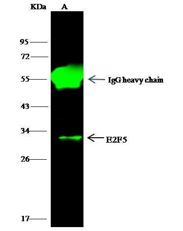
- E2F5 Immunoprecipitation using: Lane A: 0.5 mg Raji Whole Cell Lysate 1 µL with E2F5 Polyclonal Antibody (Product # PA5-81166) and 15 µL of 50 % Protein G agarose. Primary antibody: E2F5 Polyclonal Antibody, at 1:500 dilution. Secondary antibody: Dylight 800-labeled antibody to rabbit IgG (H+L), at 1:5,000 dilution. Developed using the Odyssey technique. Performed under reducing conditions. Predicted band size: 38 kDa. Observed band size: 30 kDa.
- Submitted by
- Invitrogen Antibodies (provider)
- Main image
- Experimental details
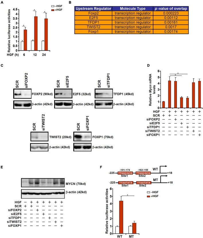
- FIGURE 2 HGF induced MYCN transcription requires an intact E2F site. (A) A MYCN promoter-luciferase construct was transfected into HepG2 cells followed by treatment with HGF. The cells were harvested at indicated time points and luciferase activities were normalized by GFP fluorescence and protein concentration. (B) IPA analysis of upstream regulators of MYCN. (C) Primary murine hepatocytes were transfected with indicated siRNAs. Knockdown efficiencies were verified by Western. (D,E) Primary murine hepatocytes were transfected with indicated siRNAs followed by treatment with HGF for 24 h. MYCN expression was examined by qPCR and Western. (F) WT or mutant MYCN promoter-luciferase construct was transfected into HepG2 cells followed by treatment with HGF for 24 h. Luciferase activities were normalized by GFP fluorescence and protein concentration. Data are expressed as mean +- standard deviation (SD). * p
- Submitted by
- Invitrogen Antibodies (provider)
- Main image

- Experimental details
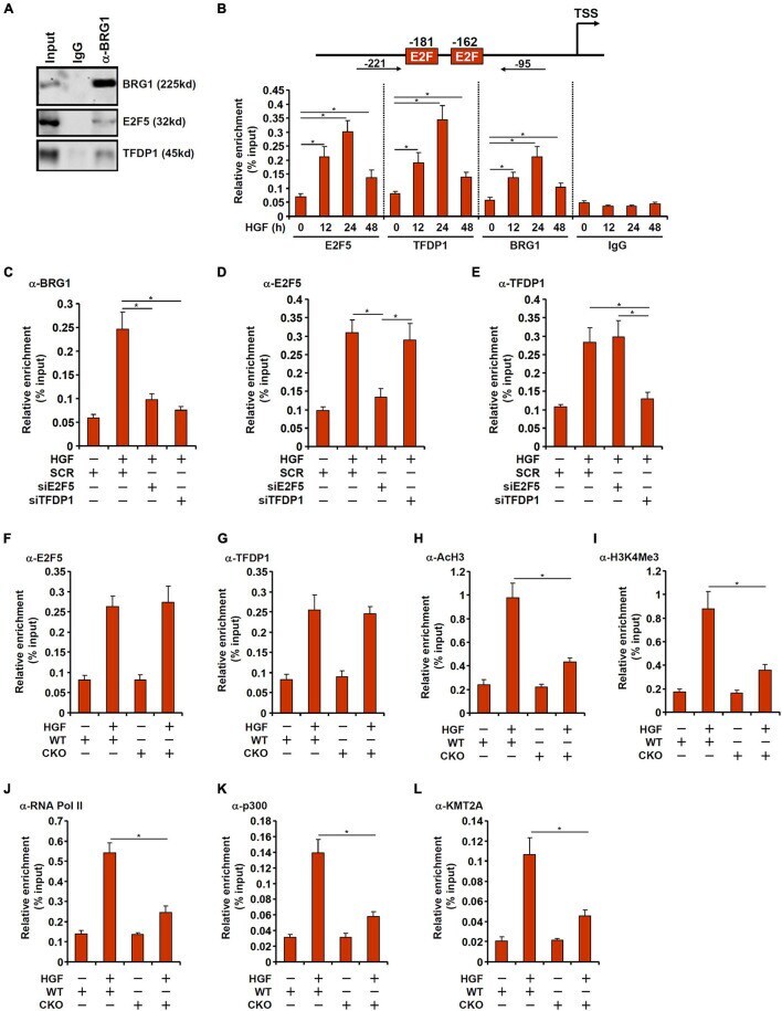
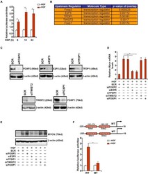
- FIGURE 4 BRG1 interacts with E2F5-TFDP1 to activate MYCN transcription. (A) Immunoprecipitation was performed with anti-BRG1 or IgG using primary murine hepatocyte lysates. (B) Primary murine hepatocytes were treated with HGF and harvested at indicated time points. ChIP assays were performed with anti-BRG1, anti-E2F5, anti-TFDP1, or IgG. Upper panel, a scheme of the MYCN promoter highlighting the positions of the two proximal E2F sites and the ChIP primers. TSS, transcription start site. (C-E) Primary murine hepatocytes were transfected with indicated siRNAs followed by treatment with HGF. ChIP assay was performed with anti-BRG1, anti-E2F5, and anti-TFDP1. (F-L) Primary hepatocytes were isolated from WT and BRG1 CKO mice followed by treatment with HGF. ChIP assays were performed with anti-E2F5, anti-TFDP1, anti-acetyl H3, anti-trimethyl H3K4, anti-RNA polymerase II, anti-p300, and anti-KMT2A. Data are expressed as mean +- standard deviation (SD). * p < 0.05 (one-way ANOVA with post hoc Scheffe test).