Explore
 Validate
 Learn
Learn Western blot
Antibody data
- Antibody Data
- Antigen structure
- References [9]
- Comments [0]
- Validations
- Western blot [5]
- Immunocytochemistry [1]
- Immunohistochemistry [1]
Submit
Validation data
Reference
Comment
Report error
- Product number
- GTX100642 - Provider product page

- Provider
- GeneTex
- Proper citation
- GeneTex Cat#GTX100642, RRID:AB_1949927
- Product name
- CD36 antibody [C1C3]
- Antibody type
- Polyclonal
- Reactivity
- Human, Mouse
- Host
- Rabbit
Submitted references Choline ameliorates cardiac hypertrophy by regulating metabolic remodelling and UPRmt through SIRT3-AMPK pathway.
Hypermethylated CD36 gene affected the progression of lung cancer.
Betulin attenuates atherosclerosis in apoE(-/-) mice by up-regulating ABCA1 and ABCG1.
A novel small molecule liver X receptor transcriptional regulator, nagilactone B, suppresses atherosclerosis in apoE-deficient mice.
Heterozygous caveolin-3 mice show increased susceptibility to palmitate-induced insulin resistance.
Fumigaclavine C activates PPARγ pathway and attenuates atherogenesis in ApoE-deficient mice.
oxLDL induces inflammatory responses in vascular smooth muscle cells via urokinase receptor association with CD36 and TLR4.
Mouse strain differences in metabolic fluxes and function of ex vivo working hearts.
CD36 participates in PrP(106-126)-induced activation of microglia.
Xu M, Xue RQ, Lu Y, Yong SY, Wu Q, Cui YL, Zuo XT, Yu XJ, Zhao M, Zang WJ
Cardiovascular research 2019 Mar 1;115(3):530-545
Cardiovascular research 2019 Mar 1;115(3):530-545
Hypermethylated CD36 gene affected the progression of lung cancer.
Sun Q, Zhang W, Wang L, Guo F, Song D, Zhang Q, Zhang D, Fan Y, Wang J
Gene 2018 Dec 15;678:395-406
Gene 2018 Dec 15;678:395-406
Betulin attenuates atherosclerosis in apoE(-/-) mice by up-regulating ABCA1 and ABCG1.
Gui YZ, Yan H, Gao F, Xi C, Li HH, Wang YP
Acta pharmacologica Sinica 2016 Sep;37(10):1337-1348
Acta pharmacologica Sinica 2016 Sep;37(10):1337-1348
A novel small molecule liver X receptor transcriptional regulator, nagilactone B, suppresses atherosclerosis in apoE-deficient mice.
Gui Y, Yao S, Yan H, Hu L, Yu C, Gao F, Xi C, Li H, Ye Y, Wang Y
Cardiovascular research 2016 Oct;112(1):502-14
Cardiovascular research 2016 Oct;112(1):502-14
Heterozygous caveolin-3 mice show increased susceptibility to palmitate-induced insulin resistance.
Talukder MA, Preda M, Ryzhova L, Prudovsky I, Pinz IM
Physiological reports 2016 Mar;4(6)
Physiological reports 2016 Mar;4(6)
Fumigaclavine C activates PPARγ pathway and attenuates atherogenesis in ApoE-deficient mice.
Du RH, Qin SY, Shi LS, Zhou ZQ, Zhu XY, Liu J, Tan RX, Cao W
Atherosclerosis 2014 May;234(1):120-8
Atherosclerosis 2014 May;234(1):120-8
oxLDL induces inflammatory responses in vascular smooth muscle cells via urokinase receptor association with CD36 and TLR4.
Kiyan Y, Tkachuk S, Hilfiker-Kleiner D, Haller H, Fuhrman B, Dumler I
Journal of molecular and cellular cardiology 2014 Jan;66:72-82
Journal of molecular and cellular cardiology 2014 Jan;66:72-82
Mouse strain differences in metabolic fluxes and function of ex vivo working hearts.
Vaillant F, Lauzier B, Poirier I, Gélinas R, Rivard ME, Robillard Frayne I, Thorin E, Des Rosiers C
American journal of physiology. Heart and circulatory physiology 2014 Jan 1;306(1):H78-87
American journal of physiology. Heart and circulatory physiology 2014 Jan 1;306(1):H78-87
CD36 participates in PrP(106-126)-induced activation of microglia.
Kouadir M, Yang L, Tan R, Shi F, Lu Y, Zhang S, Yin X, Zhou X, Zhao D
PloS one 2012;7(1):e30756
PloS one 2012;7(1):e30756
No comments: Submit comment
Supportive validation
- Submitted by
- GeneTex (provider)
- Main image
- Experimental details
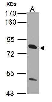
- Sample (30 ?g of whole cell lysate) A: K562 10% SDS PAGE GTX100642 diluted at 1:1000 The HRP-conjugated anti-rabbit IgG antibody (GTX213110-01) was used to detect the primary antibody.
- Submitted by
- GeneTex (provider)
- Main image
- Experimental details
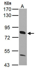
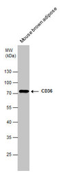
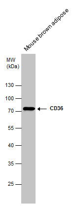
- Sample (50 ug of whole cell lysate) A: Mouse brown adipose 7.5% SDS PAGE GTX100642 diluted at 1:1000
- Validation comment
- WB
- Submitted by
- GeneTex (provider)
- Main image
- Experimental details
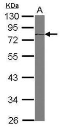
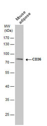
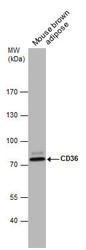
- CD36 antibody detects CD36 protein by western blot analysis. Mouse tissue extracts (50 ?g) was separated by 10 % SDS-PAGE, and the membrane was blotted with CD36 antibody (GTX100642) diluted by 1:1000.
- Validation comment
- WB
- Submitted by
- GeneTex (provider)
- Main image
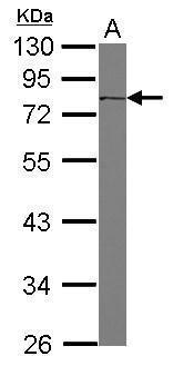
- Experimental details
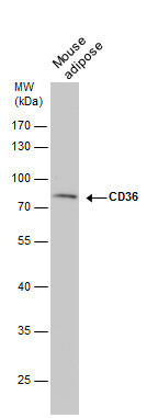
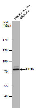
- CD36 antibody detects CD36 protein by western blot analysis. Mouse tissue extracts (50 ?g) was separated by 10% SDS-PAGE, and the membrane was blotted with CD36 antibody (GTX100642) diluted at 1:1000. The HRP-conjugated anti-rabbit IgG antibody (GTX213110-01) was used to detect the primary antibody.
- Submitted by
- GeneTex (provider)
- Main image
- Experimental details
- Mouse tissue extract (50 £gg) was separated by 7.5% SDS-PAGE, and the membrane was blotted with CD36 antibody [C1C3] (GTX100642) diluted at 1:1000.
Supportive validation
- Submitted by
- GeneTex (provider)
- Main image

- Experimental details
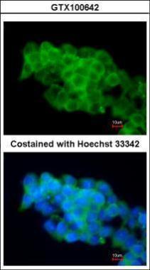
- Immunofluorescence analysis of paraformaldehyde-fixed mouse ESC D3, using CD36(GTX100642) antibody at 1:200 dilution.
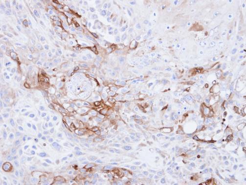
Supportive validation
- Submitted by
- GeneTex (provider)
- Main image

- Experimental details
- Immunohistochemical analysis of paraffin-embedded Ca922 xenograft, using CD36(GTX100642) antibody at 1:500 dilution.