 Explore
 Validate
 Learn
Learn Western blot
Antibody data
- Antibody Data
- Antigen structure
- References [1]
- Comments [0]
- Validations
- Western blot [3]
- Immunohistochemistry [1]
Submit
Validation data
Reference
Comment
Report error
- Product number
- MAB19552 - Provider product page

- Provider
- Novus Biologicals
- Product name
- Mouse Monoclonal CD36/SR-B3 Antibody
- Antibody type
- Monoclonal
- Description
- Protein A or G purified from hybridoma culture supernatant. Detects human CD36/SR-B3 in direct ELISAs.
- Reactivity
- Human
- Host
- Mouse
- Conjugate
- Unconjugated
- Isotype
- IgG
- Vial size
- 100 ug
- Storage
- Use a manual defrost freezer and avoid repeated freeze-thaw cycles. 12 months from date of receipt, -20 to -70 degreesC as supplied. 1 month, 2 to 8 degreesC under sterile conditions after reconstitution. 6 months, -20 to -70 degreesC under sterile conditions after reconstitution.
Submitted references Nanoparticle biointerfacing by platelet membrane cloaking.
Hu CM, Fang RH, Wang KC, Luk BT, Thamphiwatana S, Dehaini D, Nguyen P, Angsantikul P, Wen CH, Kroll AV, Carpenter C, Ramesh M, Qu V, Patel SH, Zhu J, Shi W, Hofman FM, Chen TC, Gao W, Zhang K, Chien S, Zhang L
Nature 2015 Oct 1;526(7571):118-21
Nature 2015 Oct 1;526(7571):118-21
No comments: Submit comment
Supportive validation
- Submitted by
- Novus Biologicals (provider)
- Main image

- Experimental details
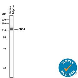
- Detection of Human CD36/SR-B3 by Western Blot. Western blot shows lysates of human placenta tissue and human platelets. PVDF membrane was probed with 1 µg/mL of Mouse Anti-Human CD36/SR-B3 Mono-clonal Antibody (Catalog # MAB19552) followed by HRP-conjugated Anti-Mouse IgG Secondary Antibody (Catalog # HAF018). A specific band was detected for CD36/SR-B3 at approximately 85-90 kDa (as indicated). This experiment was conducted under reducing conditions and using Immunoblot Buffer Group 1.

- Submitted by
- Novus Biologicals (provider)
- Main image

- Experimental details
- Detection of Human CD36/SR-B3 by Simple WesternTM. Simple Western lane view shows lysates of human platelets, loaded at 0.5 mg/mL. A specific band was detected for CD36/SR-B3 at approximately 138 kDa (as indicated) using 10 µg/mL of Mouse Anti-Human CD36/SR-B3 Monoclonal Antibody (Catalog # MAB19552) . This experiment was conducted under reducing conditions and using the 12-230 kDa separation system.

- Submitted by
- Novus Biologicals (provider)
- Main image
- Experimental details
- Detection of Human CD36/SR-B3 by Simple WesternTM. Detection of Human CD36/SR-B3 by Simple WesternTM.

Supportive validation
- Submitted by
- Novus Biologicals (provider)
- Main image
- Experimental details
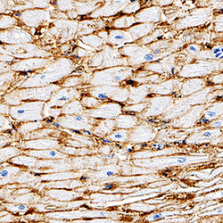
- CD36/SR-B3 in Human Heart. CD36/SR-B3 was detected in immersion fixed paraffin-embedded sections of human heart using Mouse Anti-Human CD36/SR-B3 Monoclonal Antibody (Catalog # MAB19552) at 5 µg/mL for 1 hour at room temperature followed by incubation with Anti-Mouse IgG VisUCyte™ HRP Polymer Antibody (Catalog # VC001). Tissue was stained with DAB (brown) and counterstained with hematoxylin (blue). Specific staining was localized to plasma membrane. View our protocol for Chromogenic IHC Staining of Paraffin-embedded Tissue Sections.
