Explore
 Validate
 Learn
Learn Flow cytometry
Antibody data
- Antibody Data
- Antigen structure
- References [12]
- Comments [0]
- Validations
- Flow cytometry [1]
- Other assay [7]
Submit
Validation data
Reference
Comment
Report error
- Product number
- 11-0369-41 - Provider product page

- Provider
- Invitrogen Antibodies
- Product name
- CD36 Monoclonal Antibody (eBioNL07 (NL07)), FITC, eBioscience™
- Antibody type
- Monoclonal
- Antigen
- Other
- Description
- Description: The monoclonal antibody eBioNL07 recognizes human CD36, which is a member of the class B scavenger receptor family. CD36 was originally identified as a platelet-membrane glycoprotein also called glycoprotein IV and a receptor for thrombospondin-1 (TSP-1) and extracellular matrix proteins. Binding to TSP-1 is in the CLESH (CD36 LIMP-II Emp sequence homology) domain of CD36. CD36 expression is broad and includes microvascular (but not large vessel) endothelium, adipocytes, skeletal muscle, dendritic cells, epithelia of the retina, breast, and intestine, smooth muscle cells, and hematopoietic cells, including erythroid precursors, platelets, monocytes/macrophages, DCs and megakaryocytes. Expression on platelets is absent on Nak-a negative donors. Unlike other scavenger receptor, CD36 binds LDL that has been exposed to "minimally" oxidizing conditions. CD36 is also a fatty acid translocase (FAT) necessary for the transport of long-chain fatty acids (LCFAs) and therefore may play a role in atherosclerosis. Applications Reported: This eBioNL07 (NL07) antibody has been reported for use in flow cytometric analysis. Applications Tested: This eBioNL07 (NL07) antibody has been pre-titrated and tested by flow cytometric analysis. This can be used at 5 µL (0.25 µg) per test. A test is defined as the amount (µg) of antibody that will stain a cell sample in a final volume of 100 µL. Cell number should be determined empirically but can range from 10^5 to 10^8 cells/test. Excitation: 488 nm; Emission: 520 nm; Laser: Blue Laser. Filtration: 0.2 µm post-manufacturing filtered.
- Reactivity
- Human
- Host
- Mouse
- Conjugate
- Green dye
- Isotype
- IgM
- Antibody clone number
- eBioNL07 (NL07)
- Vial size
- 25 Tests
- Concentration
- 5 µL/Test
- Storage
- 4° C, store in dark, DO NOT FREEZE!
Submitted references Genome-Wide Transcriptional Regulation of the Long Non-coding RNA Steroid Receptor RNA Activator in Human Erythroblasts.
Single-Cell Analyses Reveal Megakaryocyte-Biased Hematopoiesis in Myelofibrosis and Identify Mutant Clone-Specific Targets.
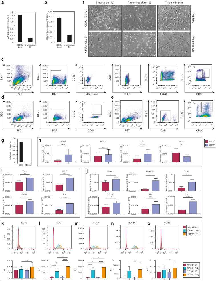
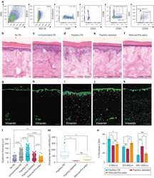
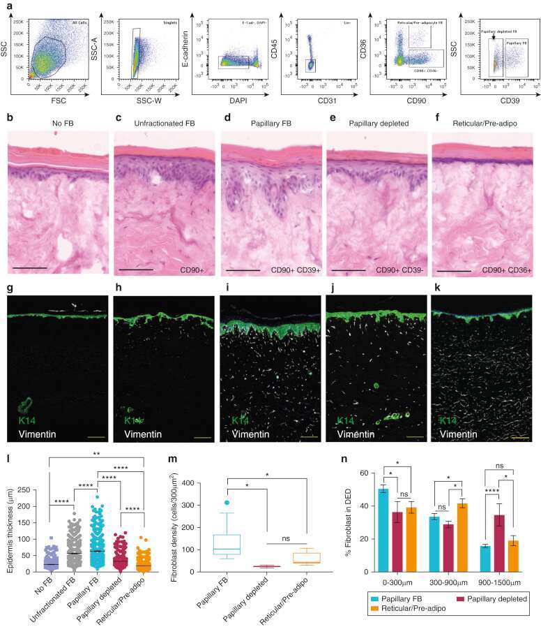
Spatial and Single-Cell Transcriptional Profiling Identifies Functionally Distinct Human Dermal Fibroblast Subpopulations.
Interferon-γ enhances both the anti-bacterial and the pro-inflammatory response of human mast cells to Staphylococcus aureus.
Developmental transcriptome analysis of human erythropoiesis.
IRGM1 regulates oxidized LDL uptake by macrophage via actin-dependent receptor internalization during atherosclerosis.
Terminal differentiation and loss of tumorigenicity of human cancers via pluripotency-based reprogramming.
CpG and non-CpG oligodeoxynucleotides directly costimulate mouse and human CD4+ T cells through a TLR9- and MyD88-independent mechanism.
An unrestrained proinflammatory M1 macrophage population induced by iron impairs wound healing in humans and mice.
Methionine 156 in the immunodominant domain of CD36 contributes to define the epitope recognized by the NL07 MoAb.
Platelet activation and inhibition of malarial cytoadherence by the anti-CD36 IgM monoclonal antibody NL07.
Analysis of the human CD36 leucocyte differentiation antigen by means of the monoclonal antibody NL07.
Sawaengdee W, Cui K, Zhao K, Hongeng S, Fucharoen S, Wongtrakoongate P
Frontiers in genetics 2020;11:850
Frontiers in genetics 2020;11:850
Single-Cell Analyses Reveal Megakaryocyte-Biased Hematopoiesis in Myelofibrosis and Identify Mutant Clone-Specific Targets.
Psaila B, Wang G, Rodriguez-Meira A, Li R, Heuston EF, Murphy L, Yee D, Hitchcock IS, Sousos N, O'Sullivan J, Anderson S, Senis YA, Weinberg OK, Calicchio ML, NIH Intramural Sequencing Center, Iskander D, Royston D, Milojkovic D, Roberts I, Bodine DM, Thongjuea S, Mead AJ
Molecular cell 2020 May 7;78(3):477-492.e8
Molecular cell 2020 May 7;78(3):477-492.e8
Spatial and Single-Cell Transcriptional Profiling Identifies Functionally Distinct Human Dermal Fibroblast Subpopulations.
Philippeos C, Telerman SB, Oulès B, Pisco AO, Shaw TJ, Elgueta R, Lombardi G, Driskell RR, Soldin M, Lynch MD, Watt FM
The Journal of investigative dermatology 2018 Apr;138(4):811-825
The Journal of investigative dermatology 2018 Apr;138(4):811-825
Interferon-γ enhances both the anti-bacterial and the pro-inflammatory response of human mast cells to Staphylococcus aureus.
Swindle EJ, Brown JM, Rådinger M, DeLeo FR, Metcalfe DD
Immunology 2015 Nov;146(3):470-85
Immunology 2015 Nov;146(3):470-85
Developmental transcriptome analysis of human erythropoiesis.
Shi L, Lin YH, Sierant MC, Zhu F, Cui S, Guan Y, Sartor MA, Tanabe O, Lim KC, Engel JD
Human molecular genetics 2014 Sep 1;23(17):4528-42
Human molecular genetics 2014 Sep 1;23(17):4528-42
IRGM1 regulates oxidized LDL uptake by macrophage via actin-dependent receptor internalization during atherosclerosis.
Xia F, Li R, Wang C, Yang S, Tian L, Dong H, Pei C, He S, Jiang P, Cheng H, Fang S, Li H, Xu H
Scientific reports 2013;3:1867
Scientific reports 2013;3:1867
Terminal differentiation and loss of tumorigenicity of human cancers via pluripotency-based reprogramming.
Zhang X, Cruz FD, Terry M, Remotti F, Matushansky I
Oncogene 2013 May 2;32(18):2249-60, 2260.e1-21
Oncogene 2013 May 2;32(18):2249-60, 2260.e1-21
CpG and non-CpG oligodeoxynucleotides directly costimulate mouse and human CD4+ T cells through a TLR9- and MyD88-independent mechanism.
Landrigan A, Wong MT, Utz PJ
Journal of immunology (Baltimore, Md. : 1950) 2011 Sep 15;187(6):3033-43
Journal of immunology (Baltimore, Md. : 1950) 2011 Sep 15;187(6):3033-43
An unrestrained proinflammatory M1 macrophage population induced by iron impairs wound healing in humans and mice.
Sindrilaru A, Peters T, Wieschalka S, Baican C, Baican A, Peter H, Hainzl A, Schatz S, Qi Y, Schlecht A, Weiss JM, Wlaschek M, Sunderkötter C, Scharffetter-Kochanek K
The Journal of clinical investigation 2011 Mar;121(3):985-97
The Journal of clinical investigation 2011 Mar;121(3):985-97
Methionine 156 in the immunodominant domain of CD36 contributes to define the epitope recognized by the NL07 MoAb.
Gruarin P, Ulliers D, Thorne RF, Alessio M
Molecular and cellular biochemistry 2000 Nov;214(1-2):89-95
Molecular and cellular biochemistry 2000 Nov;214(1-2):89-95
Platelet activation and inhibition of malarial cytoadherence by the anti-CD36 IgM monoclonal antibody NL07.
Alessio M, Greco NJ, Primo L, Ghigo D, Bosia A, Tandon NN, Ockenhouse CF, Jamieson GA, Malavasi F
Blood 1993 Dec 15;82(12):3637-47
Blood 1993 Dec 15;82(12):3637-47
Analysis of the human CD36 leucocyte differentiation antigen by means of the monoclonal antibody NL07.
Alessio M, Ghigo D, Garbarino G, Geuna M, Malavasi F
Cellular immunology 1991 Oct 15;137(2):487-500
Cellular immunology 1991 Oct 15;137(2):487-500
No comments: Submit comment
Supportive validation
- Submitted by
- Invitrogen Antibodies (provider)
- Main image
- Experimental details
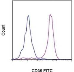
- Staining of normal human peripheral blood cells with Mouse IgM Isotype Control FITC (Product # 11-4752-80) (blue histogram) or Anti-Human CD36 FITC (purple histogram). Cells in the monocyte gate were used for analysis.
- Conjugate
- Green dye

Supportive validation
- Submitted by
- Invitrogen Antibodies (provider)
- Main image
- Experimental details
- NULL
- Conjugate
- Green dye

- Submitted by
- Invitrogen Antibodies (provider)
- Main image
- Experimental details
- NULL
- Conjugate
- Green dye
- Submitted by
- Invitrogen Antibodies (provider)
- Main image
- Experimental details
- NULL
- Conjugate
- Green dye
- Submitted by
- Invitrogen Antibodies (provider)
- Main image
- Experimental details
- NULL
- Conjugate
- Green dye

- Submitted by
- Invitrogen Antibodies (provider)
- Main image
- Experimental details
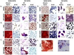
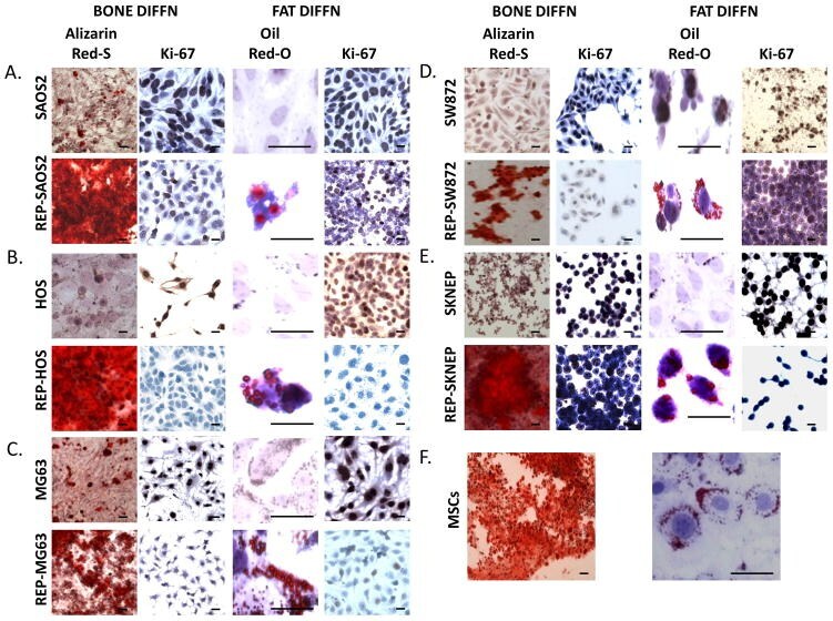
- Figure 2 (A-E) Comparison of sarcomas and reprogrammed-sarcomas in terms of achieving terminal differentiation as measured by acquisition of the terminal phenotype (both bone via Alizarin Red S staining for calcium deposition and fat via Oil-Red-O staining for lipid accumulation) AND cessation of proliferation via loss of Ki67. (F) MSCs differentiated as controls into bone and fat. (Oil-Red-O images are magnified 10-fold to accentuate lipid formation associated with each cell). Scale bars=10um.
- Conjugate
- Green dye
- Submitted by
- Invitrogen Antibodies (provider)
- Main image
- Experimental details
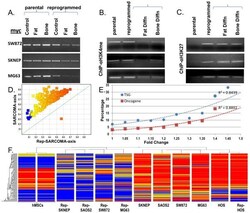
- Figure 4 (A) Myc RT-PCR in parental and reprogrammed sarcoma cells following three weeks in either maintenance (control), adipogenic (fat) or osteogenic (bone) differentiation medium. Chromatin immunoprecipitation of the myc promoter using anti-H3K4triMe (B) or anti-HEK27triMe (C) antibodies in either maintenance (control), adipogenic (fat) or osteogenic (bone) differentiation medium. (D) DNA-promoter methylation analysis showing all statistically differentially methylated promoters (T-Test p
- Conjugate
- Green dye
- Submitted by
- Invitrogen Antibodies (provider)
- Main image
- Experimental details
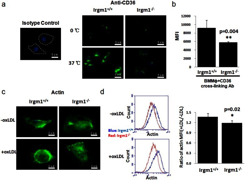
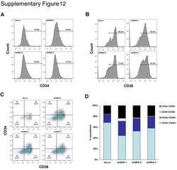
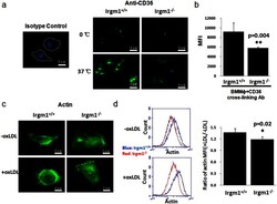
- Figure 4 IRGM1 controls CD36 internalization by regulating actin polymerization. BMMPhi from either Irgm1 +/+ or Irgm1 -/- mice was labeled with CD36 cross-linking antibody. Then, they were either left in 37degC to cross-link CD36 or on ice. Cells were then washed by cold acid wash buffer to deplete surface CD36. Confocal microscope and flow cytometry were used to detect and quantify CD36 internalization respectively. (a and b n = 8/group, p = 0.004). F-actin polymerization was detected by phalloidin-FITC in BMMPhi from either Irgm1 +/+ or Irgm1 -/- mice with or without oxLDL. The fluorescence was measured by confocal microscope and quantified by flow cytometry (c and d, n = 3, p = 0.02). Data are represented as mean +- SEM.
- Conjugate
- Green dye