Explore
 Validate
 Learn
Learn Flow cytometry
Antibody data
- Antibody Data
- Antigen structure
- References [8]
- Comments [0]
- Validations
- Flow cytometry [1]
- Other assay [4]
Submit
Validation data
Reference
Comment
Report error
- Product number
- 46-0369-41 - Provider product page

- Provider
- Invitrogen Antibodies
- Product name
- CD36 Monoclonal Antibody (eBioNL07 (NL07)), PerCP-eFluor™ 710, eBioscience™
- Antibody type
- Monoclonal
- Antigen
- Other
- Description
- Description: The monoclonal antibody eBioNL07 recognizes human CD36, which is a member of the class B scavenger receptor family. CD36 was originally identified as a platelet-membrane glycoprotein also called glycoprotein IV and a receptor for thrombospondin-1 (TSP-1) and extracellular matrix proteins. Binding to TSP-1 is in the CLESH (CD36 LIMP-II Emp sequence homology) domain of CD36. CD36 expression is broad and includes microvascular (but not large vessel) endothelium, adipocytes, skeletal muscle, dendritic cells, epithelia of the retina, breast, and intestine, smooth muscle cells, and hematopoietic cells, including erythroid precursors, platelets, monocytes/macrophages, DCs and megakaryocytes. Expression on platelets is absent on Nak-a negative donors. Unlike other scavenger receptor, CD36 binds LDL that has been exposed to "minimally" oxidizing conditions. CD36 is also a fatty acid translocase (FAT) necessary for the transport of long-chain fatty acids (LCFAs) and therefore may play a role in atherosclerosis. Applications Reported: This eBioNL07 (NL07) antibody has been reported for use in flow cytometric analysis. Applications Tested: This eBioNL07 (NL07) antibody has been pre-titrated and tested by flow cytometric analysis of normal human peripheral bloocd cells. This can be used at 5 µL (0.125 µg) per test. A test is defined as the amount (µg) of antibody that will stain a cell sample in a final volume of 100 µL. Cell number should be determined empirically but can range from 10^5 to 10^8 cells/test. PerCP-eFluor® 710 emits at 710 nm and is excited with the blue laser (488 nm); it can be used in place of PerCP-Cyanine5.5. We recommend using a 710/50 bandpass filter, however, the 695/40 bandpass filter is an acceptable alternative. Please make sure that your instrument is capable of detecting this fluorochrome. Fixation: Samples can be stored in IC Fixation Buffer (Product # 00-822-49) (100 µL cell sample + 100 µL IC Fixation Buffer) or 1-step Fix/Lyse Solution (Product # 00-5333-54) for up to 3 days in the dark at 4°C with minimal impact on brightness and FRET efficiency/compensation. Some generalizations regarding fluorophore performance after fixation can be made, but clone specific performance should be determined empirically. Excitation: 488 nm; Emission: 710 nm; Laser: Blue Laser. Filtration: 0.2 µm post-manufacturing filtered.
- Reactivity
- Human
- Host
- Mouse
- Isotype
- IgM
- Antibody clone number
- eBioNL07 (NL07)
- Vial size
- 25 Tests
- Concentration
- 5 µL/Test
- Storage
- 4° C, store in dark, DO NOT FREEZE!
Submitted references Genome-Wide Transcriptional Regulation of the Long Non-coding RNA Steroid Receptor RNA Activator in Human Erythroblasts.
Lymph protects metastasizing melanoma cells from ferroptosis.
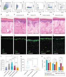
Spatial and Single-Cell Transcriptional Profiling Identifies Functionally Distinct Human Dermal Fibroblast Subpopulations.
CD36-mediated hematoma absorption following intracerebral hemorrhage: negative regulation by TLR4 signaling.
IRGM1 regulates oxidized LDL uptake by macrophage via actin-dependent receptor internalization during atherosclerosis.
Methionine 156 in the immunodominant domain of CD36 contributes to define the epitope recognized by the NL07 MoAb.
Platelet activation and inhibition of malarial cytoadherence by the anti-CD36 IgM monoclonal antibody NL07.
Analysis of the human CD36 leucocyte differentiation antigen by means of the monoclonal antibody NL07.
Sawaengdee W, Cui K, Zhao K, Hongeng S, Fucharoen S, Wongtrakoongate P
Frontiers in genetics 2020;11:850
Frontiers in genetics 2020;11:850
Lymph protects metastasizing melanoma cells from ferroptosis.
Ubellacker JM, Tasdogan A, Ramesh V, Shen B, Mitchell EC, Martin-Sandoval MS, Gu Z, McCormick ML, Durham AB, Spitz DR, Zhao Z, Mathews TP, Morrison SJ
Nature 2020 Sep;585(7823):113-118
Nature 2020 Sep;585(7823):113-118
Spatial and Single-Cell Transcriptional Profiling Identifies Functionally Distinct Human Dermal Fibroblast Subpopulations.
Philippeos C, Telerman SB, Oulès B, Pisco AO, Shaw TJ, Elgueta R, Lombardi G, Driskell RR, Soldin M, Lynch MD, Watt FM
The Journal of investigative dermatology 2018 Apr;138(4):811-825
The Journal of investigative dermatology 2018 Apr;138(4):811-825
CD36-mediated hematoma absorption following intracerebral hemorrhage: negative regulation by TLR4 signaling.
Fang H, Chen J, Lin S, Wang P, Wang Y, Xiong X, Yang Q
Journal of immunology (Baltimore, Md. : 1950) 2014 Jun 15;192(12):5984-92
Journal of immunology (Baltimore, Md. : 1950) 2014 Jun 15;192(12):5984-92
IRGM1 regulates oxidized LDL uptake by macrophage via actin-dependent receptor internalization during atherosclerosis.
Xia F, Li R, Wang C, Yang S, Tian L, Dong H, Pei C, He S, Jiang P, Cheng H, Fang S, Li H, Xu H
Scientific reports 2013;3:1867
Scientific reports 2013;3:1867
Methionine 156 in the immunodominant domain of CD36 contributes to define the epitope recognized by the NL07 MoAb.
Gruarin P, Ulliers D, Thorne RF, Alessio M
Molecular and cellular biochemistry 2000 Nov;214(1-2):89-95
Molecular and cellular biochemistry 2000 Nov;214(1-2):89-95
Platelet activation and inhibition of malarial cytoadherence by the anti-CD36 IgM monoclonal antibody NL07.
Alessio M, Greco NJ, Primo L, Ghigo D, Bosia A, Tandon NN, Ockenhouse CF, Jamieson GA, Malavasi F
Blood 1993 Dec 15;82(12):3637-47
Blood 1993 Dec 15;82(12):3637-47
Analysis of the human CD36 leucocyte differentiation antigen by means of the monoclonal antibody NL07.
Alessio M, Ghigo D, Garbarino G, Geuna M, Malavasi F
Cellular immunology 1991 Oct 15;137(2):487-500
Cellular immunology 1991 Oct 15;137(2):487-500
No comments: Submit comment
Supportive validation
- Submitted by
- Invitrogen Antibodies (provider)
- Main image
- Experimental details
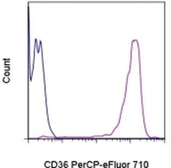
- Staining of normal human peripheral blood cells with staining buffer (autofluorescence) (blue histogram) or Anti-Human CD36 PerCP-eFluor® 710 (purple histogram). Cells in the monocyte gate were used for analysis.

Supportive validation
- Submitted by
- Invitrogen Antibodies (provider)
- Main image
- Experimental details
- NULL
- Submitted by
- Invitrogen Antibodies (provider)
- Main image
- Experimental details
- NULL
- Submitted by
- Invitrogen Antibodies (provider)
- Main image
- Experimental details
- NULL
- Submitted by
- Invitrogen Antibodies (provider)
- Main image
- Experimental details
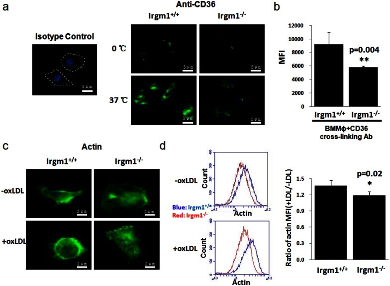
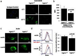
- Figure 4 IRGM1 controls CD36 internalization by regulating actin polymerization. BMMPhi from either Irgm1 +/+ or Irgm1 -/- mice was labeled with CD36 cross-linking antibody. Then, they were either left in 37degC to cross-link CD36 or on ice. Cells were then washed by cold acid wash buffer to deplete surface CD36. Confocal microscope and flow cytometry were used to detect and quantify CD36 internalization respectively. (a and b n = 8/group, p = 0.004). F-actin polymerization was detected by phalloidin-FITC in BMMPhi from either Irgm1 +/+ or Irgm1 -/- mice with or without oxLDL. The fluorescence was measured by confocal microscope and quantified by flow cytometry (c and d, n = 3, p = 0.02). Data are represented as mean +- SEM.