Explore
 Validate
 Learn
Learn Western blot
Antibody data
- Antibody Data
- Antigen structure
- References [3]
- Comments [0]
- Validations
- Western blot [4]
- Immunocytochemistry [1]
- Immunohistochemistry [3]
- Other assay [2]
Submit
Validation data
Reference
Comment
Report error
- Product number
- PA5-27236 - Provider product page

- Provider
- Invitrogen Antibodies
- Product name
- CD36 Polyclonal Antibody
- Antibody type
- Polyclonal
- Antigen
- Synthetic peptide
- Description
- Recommended positive controls: K562, mouse brown adipose, RAW264.7. Store product as a concentrated solution. Centrifuge briefly prior to opening the vial.
- Reactivity
- Human, Mouse
- Host
- Rabbit
- Isotype
- IgG
- Vial size
- 100 μL
- Concentration
- 0.47 mg/mL
- Storage
- Store at 4°C short term. For long term storage, store at -20°C, avoiding freeze/thaw cycles.
Submitted references Tart Cherry Juice and Seeds Affect Pro-Inflammatory Markers in Visceral Adipose Tissue of High-Fat Diet Obese Rats.
Receptor Heterodimerization and Co-Receptor Engagement in TLR2 Activation Induced by MIC1 and MIC4 from Toxoplasma gondii.
Osteogenic Potential of Caspases Related to Endochondral Ossification.
Moruzzi M, Klöting N, Blüher M, Martinelli I, Tayebati SK, Gabrielli MG, Roy P, Micioni Di Bonaventura MV, Cifani C, Lupidi G, Amenta F, Tomassoni D
Molecules (Basel, Switzerland) 2021 Mar 5;26(5)
Molecules (Basel, Switzerland) 2021 Mar 5;26(5)
Receptor Heterodimerization and Co-Receptor Engagement in TLR2 Activation Induced by MIC1 and MIC4 from Toxoplasma gondii.
Costa Mendonça-Natividade F, Duque Lopes C, Ricci-Azevedo R, Sardinha-Silva A, Figueiredo Pinzan C, Paiva Alegre-Maller AC, L Nohara L, B Carneiro A, Panunto-Castelo A, C Almeida I, Roque-Barreira MC
International journal of molecular sciences 2019 Oct 10;20(20)
International journal of molecular sciences 2019 Oct 10;20(20)
Osteogenic Potential of Caspases Related to Endochondral Ossification.
Janečková E, Bíliková P, Matalová E
The journal of histochemistry and cytochemistry : official journal of the Histochemistry Society 2018 Jan;66(1):47-58
The journal of histochemistry and cytochemistry : official journal of the Histochemistry Society 2018 Jan;66(1):47-58
No comments: Submit comment
Supportive validation
- Submitted by
- Invitrogen Antibodies (provider)
- Main image
- Experimental details
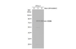
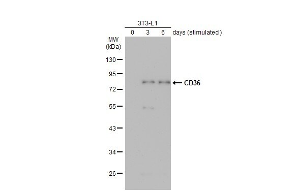
- Western Blot using CD36 Polyclonal Antibody (Product # PA5-27236). Unstimulatd and stimulatd 3T3-L1 whole cell extracts (20 µg) were separated by 10% SDS-PAGE, and the membrane was blotted with CD36 Polyclonal Antibody (Product # PA5-27236) diluted at 1:500. The HRP-conjugated anti-rabbit IgG antibody was used to detect the primary antibody. (The differentiation stimulated medium is composed by basal medium, 10% FBS, 50 µg/mL gentamicin, 1 nM L-glutamin, 500 µM IBMX, 1 µM dexamethasone, 2 µM rosiglitazone and 1 µg/mL insulin.).
- Submitted by
- Invitrogen Antibodies (provider)
- Main image
- Experimental details
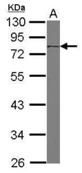
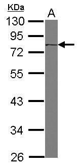
- Western Blot using CD36 Polyclonal Antibody (Product # PA5-27236). Sample (30 µg of whole cell lysate). Lane A: K562. 10% SDS PAGE. CD36 Polyclonal Antibody (Product # PA5-27236) diluted at 1:1,000. The HRP-conjugated anti-rabbit IgG antibody was used to detect the primary antibody.
- Submitted by
- Invitrogen Antibodies (provider)
- Main image
- Experimental details
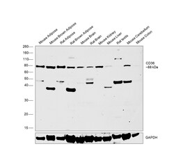
- Western Blot was performed using Anti-CD36 Polyclonal Antibody (Product # PA5-27236) and a 88 kDa band corresponding to Platelet glycoprotein 4 was observed across tissues tested and the expression was found to be higher in adipose and brown adipose tissues in comparison to other tissues tested. Tissue extracts (30 µg lysate) of Mouse Adipose (Lane 1), Mouse Brown Adipose (Lane 2), Rat Adipose (Lane 3), Rat Brown Adipose (Lane 4), Mouse Brain (Lane 5), Rat Brain (Lane 6), Mouse Kidney (Lane 7), Mouse Liver (Lane 8), Rat Testis (Lane 9), Mouse Cerebellum (Lane 10), Mouse Colon (Lane 11) were electrophoresed using NuPAGE™ 4-12% Bis-Tris Protein Gel (Product # NP0322BOX). Resolved proteins were then transferred onto a Nitrocellulose membrane (Product # IB23001) by iBlot® 2 Dry Blotting System (Product # IB21001). The Blot was probed with the primary antibody (1:1000 dilution) and detected by chemiluminescence with Goat anti-Rabbit IgG (Heavy Chain) Superclonal™ Recombinant Secondary Antibody, HRP (Product # A27036, 1:6000 dilution) using the iBright FL 1000 (Product # A32752). Chemiluminescent detection was performed using Novex® ECL Chemiluminescent Substrate Reagent Kit (Product # WP20005). Additional uncharacterized bands were also observed between 30-50 kDa.
- Submitted by
- Invitrogen Antibodies (provider)
- Main image
- Experimental details
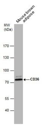
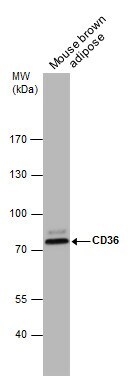
- Western Blot analysis of CD36 was performed by separating 50 µg of Mouse tissue extracts by 7.5% SDS-PAGE. Proteins were transferred to a membrane and probed with a CD36 Polyclonal Antibody (Product # PA5-27236) at a dilution of 1:1000.
Supportive validation
- Submitted by
- Invitrogen Antibodies (provider)
- Main image
- Experimental details
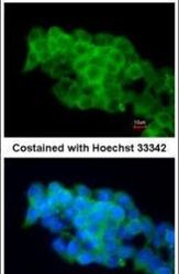
- Immunofluorescent analysis of CD36 in paraformaldehyde-fixed mouse ESC D3 cells using a CD36 polyclonal antibody (Product # PA5-27236) at a 1:200 dilution.

Supportive validation
- Submitted by
- Invitrogen Antibodies (provider)
- Main image
- Experimental details
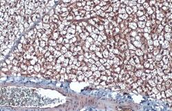
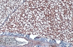
- CD36 Polyclonal Antibody detects CD36 protein by immunohistochemical analysis. Sample: Paraffin-embedded mouse brown adipocyte. CD36 stained by CD36 Polyclonal Antibody (Product # PA5-27236) diluted at 1:500. Antigen Retrieval: Citrate buffer, pH 6.0, 15 min.
- Submitted by
- Invitrogen Antibodies (provider)
- Main image
- Experimental details
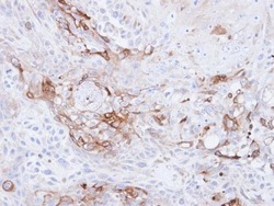
- Immunohistochemical analysis of paraffin-embedded Ca922 xenograft, using CD36 (Product # PA5-27236) antibody at 1:500 dilution. Antigen Retrieval: EDTA based buffer, pH 8.0, 15 min.

- Submitted by
- Invitrogen Antibodies (provider)
- Main image
- Experimental details
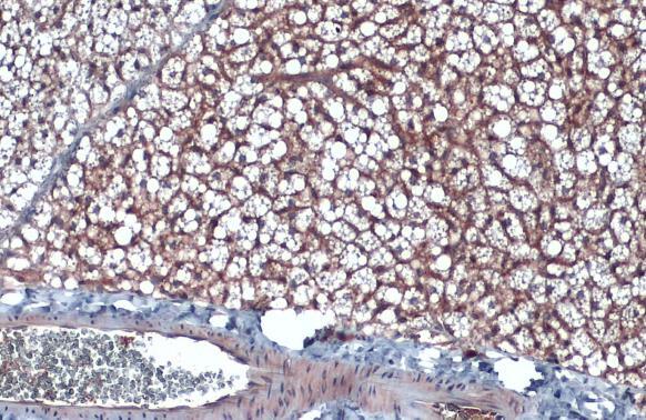
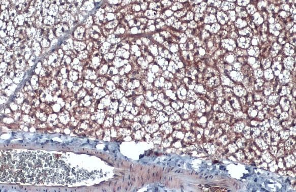
- CD36 Polyclonal Antibody detects CD36 protein by immunohistochemical analysis. Sample: Paraffin-embedded mouse brown adipocyte. CD36 stained by CD36 Polyclonal Antibody (Product # PA5-27236) diluted at 1:500. Antigen Retrieval: Citrate buffer, pH 6.0, 15 min.
Supportive validation
- Submitted by
- Invitrogen Antibodies (provider)
- Main image
- Experimental details
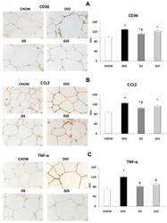
- Figure 8 Immunohistochemical analysis of RPW processed with different antibodies: ( A ) anti-CD36, ( B ) anti-CCL2 and ( C ) anti-TNF-alpha. CHOW: Standard diet; DIO: High-Fat diet; DS: High-Fat diet + Seeds; DJS: High-Fat diet + Juice and Seeds. The densitometric analysis is expressed as an arbitrary optical density unit (ODU). Data are the mean +- SEM. * p < 0.05 vs. CHOW rats; # p < 0.05 vs. DIO rats.

- Submitted by
- Invitrogen Antibodies (provider)
- Main image
- Experimental details
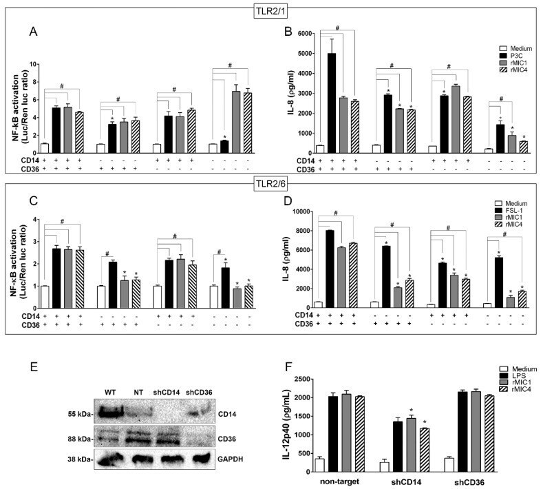
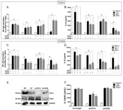
- The CD14 and CD36 co-receptors amplify TLR2-mediated cell activation induced by microneme proteins. HEK293T cells transfected with (A,B) TLR2/1 or (C,D) TLR2/6 were co-transfected with CD14 and CD36 either together or alone, along with an NF-κB-dependent luciferase reporter construct (pELAM-firefly luciferase) and a Renilla luciferase reporter construct (an internal control). The total amount of DNA in each transfection was kept constant by adding empty vector. After 48 h of transfection, the cells were stimulated with rMIC1 (50 nM) or rMIC4 (50 nM). Pam3CSK4 (P3C, 1 nM) was used as a positive control for TLR2/1, and FSL-1 (1 nM) was used as a positive control for TLR2/6. Medium was used as the negative control. Twenty-four hours post-stimulation, the IL-8 concentration in cell supernatants were assessed by ELISA. Statistical analysis was performed to compare (*) the responses in cells lacking one or more co-receptor to the responses when both CD14 and CD36 were expressed. The responses in cells expressing one co-receptor, both co-receptors, or no co-receptor (#) were compared after stimulation with microneme protein versus the responses in the negative control cells (medium). The data are from three independent experiments yielding similar results (E) BMDMs obtained from wild type C57BL/6 mice were transduced with lentivirus vectors encoding shRNA sequences for CD14 and CD36 or a non-target control shRNA. The expression levels of CD14 and CD36 were evaluated by immunoblottin