Explore
 Validate
 Learn
Learn Western blot
Antibody data
- Antibody Data
- Antigen structure
- References [5]
- Comments [0]
- Validations
- Western blot [3]
Submit
Validation data
Reference
Comment
Report error
- Product number
- AF1955 - Provider product page

- Provider
- R&D Systems
- Product name
- Human CD36/SR-B3 Antibody
- Antibody type
- Polyclonal
- Description
- Antigen Affinity-purified. Detects human CD36 in direct ELISAs and Western blots. In direct ELISAs, less than 20% cross-reactivity with recombinant mouse CD36 is observed.
- Reactivity
- Human
- Host
- Goat
- Conjugate
- Unconjugated
- Antigen sequence
P16671- Isotype
- IgG
- Vial size
- 100 ug
- Storage
- Use a manual defrost freezer and avoid repeated freeze-thaw cycles. 12 months from date of receipt, -20 to -70 °C as supplied. 1 month, 2 to 8 °C under sterile conditions after reconstitution. 6 months, -20 to -70 °C under sterile conditions after reconstitution.
Submitted references Adipocyte-induced CD36 expression drives ovarian cancer progression and metastasis.
Regulation of AMPK activation by CD36 links fatty acid uptake to β-oxidation.
A novel ELISA for measuring CD36 protein in human adipose tissue.
Preventive effects of heregulin-beta1 on macrophage foam cell formation and atherosclerosis.
Internal mammary artery smooth muscle cells resist migration and possess high antioxidant capacity.
Ladanyi A, Mukherjee A, Kenny HA, Johnson A, Mitra AK, Sundaresan S, Nieman KM, Pascual G, Benitah SA, Montag A, Yamada SD, Abumrad NA, Lengyel E
Oncogene 2018 Apr;37(17):2285-2301
Oncogene 2018 Apr;37(17):2285-2301
Regulation of AMPK activation by CD36 links fatty acid uptake to β-oxidation.
Samovski D, Sun J, Pietka T, Gross RW, Eckel RH, Su X, Stahl PD, Abumrad NA
Diabetes 2015 Feb;64(2):353-9
Diabetes 2015 Feb;64(2):353-9
A novel ELISA for measuring CD36 protein in human adipose tissue.
Allred CC, Krennmayr T, Koutsari C, Zhou L, Ali AH, Jensen MD
Journal of lipid research 2011 Feb;52(2):408-15
Journal of lipid research 2011 Feb;52(2):408-15
Preventive effects of heregulin-beta1 on macrophage foam cell formation and atherosclerosis.
Xu G, Watanabe T, Iso Y, Koba S, Sakai T, Nagashima M, Arita S, Hongo S, Ota H, Kobayashi Y, Miyazaki A, Hirano T
Circulation research 2009 Aug 28;105(5):500-10
Circulation research 2009 Aug 28;105(5):500-10
Internal mammary artery smooth muscle cells resist migration and possess high antioxidant capacity.
Mahadevan VS, Campbell M, McKeown PP, Bayraktutan U
Cardiovascular research 2006 Oct 1;72(1):60-8
Cardiovascular research 2006 Oct 1;72(1):60-8
No comments: Submit comment
Supportive validation
- Submitted by
- R&D Systems (provider)
- Main image

- Experimental details
- Detection of Human CD36/SR-B3 by Western Blot. Western blot shows lysates of human placenta tissue and human platelets. PVDF membrane was probed with 1 µg/mL of Goat Anti-Human CD36/SR-B3 Antigen Affinity-purified Polyclonal Antibody (Catalog # AF1955) followed by HRP-conjugated Anti-Goat IgG Secondary Antibody (Catalog # HAF019). A specific band was detected for CD36/SR-B3 at approximately 85-90 kDa (as indicated). This experiment was conducted under reducing conditions and using Immunoblot Buffer Group 1.

- Submitted by
- R&D Systems (provider)
- Main image
- Experimental details
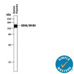
- Detection of Human CD36/SR-B3 by Simple Western<SUP abp="261">TM. Simple Western lane view shows lysates of human placenta tissue, loaded at 0.2 mg/mL. A specific band was detected for CD36/SR-B3 at approximately 140 kDa (as indicated) using 10 µg/mL of Goat Anti-Human CD36/SR-B3 Antigen Affinity-purified Polyclonal Antibody (Catalog # AF1955) followed by 1:50 dilution of HRP-conjugated Anti-Goat IgG Secondary Antibody (Catalog # HAF109). This experiment was conducted under reducing conditions and using the 12-230 kDa separation system.

- Submitted by
- R&D Systems (provider)
- Main image
- Experimental details
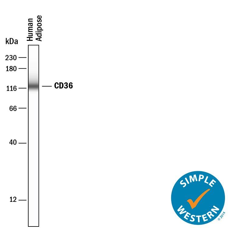
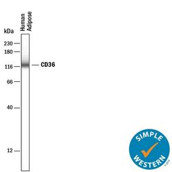
- Detection of Human CD36/SR-B3 by Simple WesternTM. Simple Western lane view shows lysates of human adipose tissue, loaded at 0.2 mg/mL. A specific band was detected for CD36/SR-B3 at approximately 125 kDa (as indicated) using 50 µg/mL of Goat Anti-Human CD36/SR-B3 Antigen Affinity-purified Polyclonal Antibody (Catalog # AF1955) followed by 1:50 dilution of HRP-conjugated Anti-Goat IgG Secondary Antibody (Catalog # HAF109). This experiment was conducted under reducing conditions and using the 12-230 kDa separation system.