Explore
 Validate
 Learn
Learn Western blot
 Immunocytochemistry
Antibody data
- Antibody Data
- Antigen structure
- References [1]
- Comments [0]
- Validations
- Immunocytochemistry [1]
- Immunohistochemistry [1]
- Other assay [1]
Submit
Validation data
Reference
Comment
Report error
- Product number
- PA5-30378 - Provider product page

- Provider
- Invitrogen Antibodies
- Product name
- IRF9 Polyclonal Antibody
- Antibody type
- Polyclonal
- Antigen
- Recombinant full-length protein
- Description
- Recommended positive controls: A549, HeLa, HepG2, HCT116. Predicted reactivity: Mouse (81%), Pig (87%), Rhesus Monkey (96%), Bovine (84%). Store product as a concentrated solution. Centrifuge briefly prior to opening the vial.
- Reactivity
- Human
- Host
- Rabbit
- Isotype
- IgG
- Vial size
- 100 μL
- Concentration
- 1.63 mg/mL
- Storage
- Store at 4°C short term. For long term storage, store at -20°C, avoiding freeze/thaw cycles.
Submitted references Modulation of epidermal transcription circuits in psoriasis: new links between inflammation and hyperproliferation.
Swindell WR, Johnston A, Xing X, Voorhees JJ, Elder JT, Gudjonsson JE
PloS one 2013;8(11):e79253
PloS one 2013;8(11):e79253
No comments: Submit comment
Supportive validation
- Submitted by
- Invitrogen Antibodies (provider)
- Main image
- Experimental details
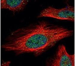
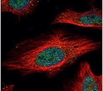
- Immunofluorescent analysis of IRF9 in paraformaldehyde-fixed HeLa cells using an IRF9 polyclonal antibody (Product # PA5-30378) (Green) at a 1:500 dilution. Alpha-tubulin filaments were labeled with Product # PA5-29281 (Red) at a 1:2000.
Supportive validation
- Submitted by
- Invitrogen Antibodies (provider)
- Main image
- Experimental details
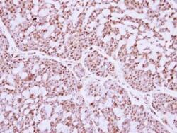
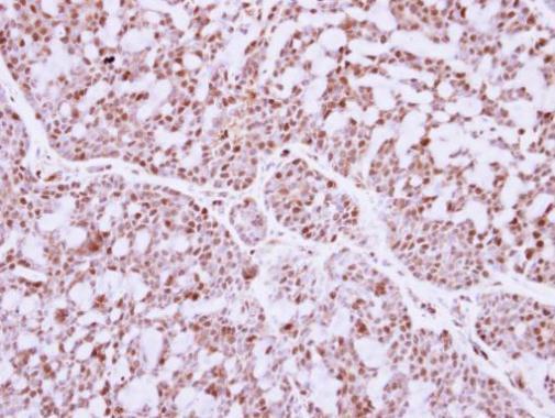
- Immunohistochemical analysis of paraffin-embedded human breast cancer, using IRF9 (Product # PA5-30378) antibody at 1:250 dilution. Antigen Retrieval: EDTA based buffer, pH 8.0, 15 min.
Supportive validation
- Submitted by
- Invitrogen Antibodies (provider)
- Main image
- Experimental details
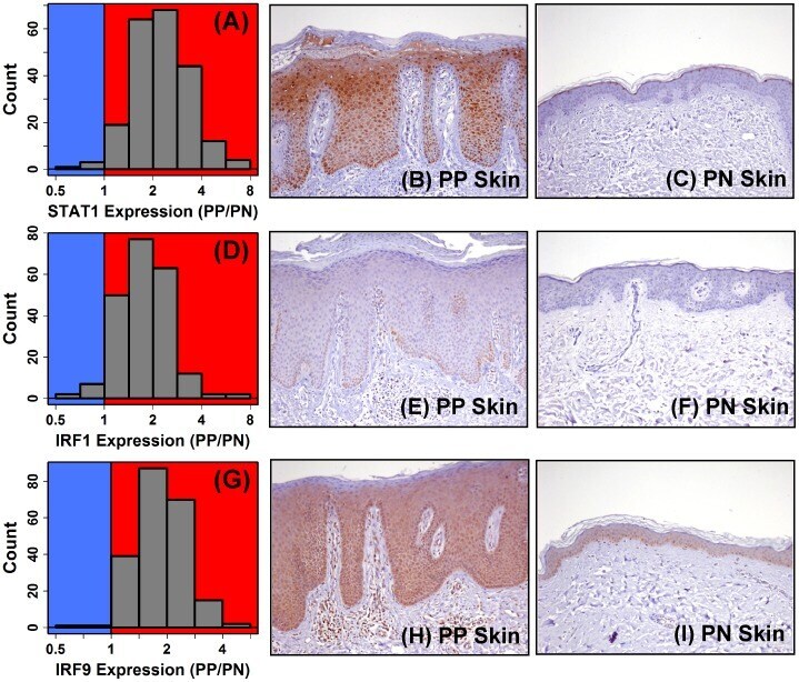
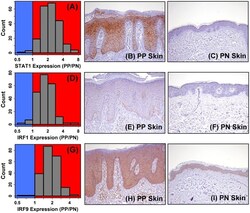
- Figure 7 STAT1 and IRF9 mRNA are elevated in psoriasis lesions with increased abundance of pSTAT1(ser727) and IRF9 protein in the psoriatic epidermis. Figures (A), (D) and (G) show the PP/PN fold-change distributions for STAT1 , IRF1 and IRF9 , respectively ( n = 215 patients). Figures (B), (E) and (H) show staining for pSTAT1(ser727), IRF1 and IRF9 in lesional (PP) skin, respectively. Figures (C), (F) and (I) show staining for pSTAT1(ser727), IRF1 and IRF9 in uninvolved (PN) skin, respectively.