 Explore
 Validate
 Learn
Learn Immunocytochemistry
Antibody data
- Antibody Data
- Antigen structure
- References [1]
- Comments [0]
- Validations
- Immunocytochemistry [2]
- Immunohistochemistry [3]
- Other assay [2]
Submit
Validation data
Reference
Comment
Report error
- Product number
- PA5-64309 - Provider product page

- Provider
- Invitrogen Antibodies
- Product name
- DNAH2 Polyclonal Antibody
- Antibody type
- Polyclonal
- Antigen
- Recombinant protein fragment
- Description
- Immunogen sequence: PGEWENACNE MQRMLIVRSL RQDRVAFCVT SFIITNLGSR FIEPPVLNMK SVLEDSTPRS PLVFILSPGV DPTSALL Highest antigen sequence identity to the following orthologs: Mouse - 96%, Rat - 95%.
- Reactivity
- Human
- Host
- Rabbit
- Isotype
- IgG
- Vial size
- 100 μL
- Concentration
- 0.1 mg/mL
- Storage
- Store at 4°C short term. For long term storage, store at -20°C, avoiding freeze/thaw cycles.
Submitted references Genetic Defects in DNAH2 Underlie Male Infertility With Multiple Morphological Abnormalities of the Sperm Flagella in Humans and Mice.
Hwang JY, Nawaz S, Choi J, Wang H, Hussain S, Nawaz M, Lopez-Giraldez F, Jeong K, Dong W, Oh JN, Bilguvar K, Mane S, Lee CK, Bystroff C, Lifton RP, Ahmad W, Chung JJ
Frontiers in cell and developmental biology 2021;9:662903
Frontiers in cell and developmental biology 2021;9:662903
No comments: Submit comment
Supportive validation
- Submitted by
- Invitrogen Antibodies (provider)
- Main image
- Experimental details
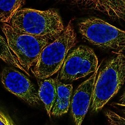
- Immunofluorescent staining of DNAH2 in human cell line SiHa using a DNAH2 Polyclonal Antibody (Product # PA5-64309) shows localization to microtubules.

- Submitted by
- Invitrogen Antibodies (provider)
- Main image

- Experimental details
- Immunofluorescent staining of DNAH2 in human cell line SiHa using a DNAH2 Polyclonal Antibody (Product # PA5-64309) shows localization to microtubules.

Supportive validation
- Submitted by
- Invitrogen Antibodies (provider)
- Main image
- Experimental details
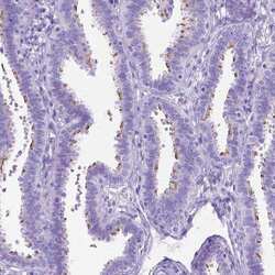
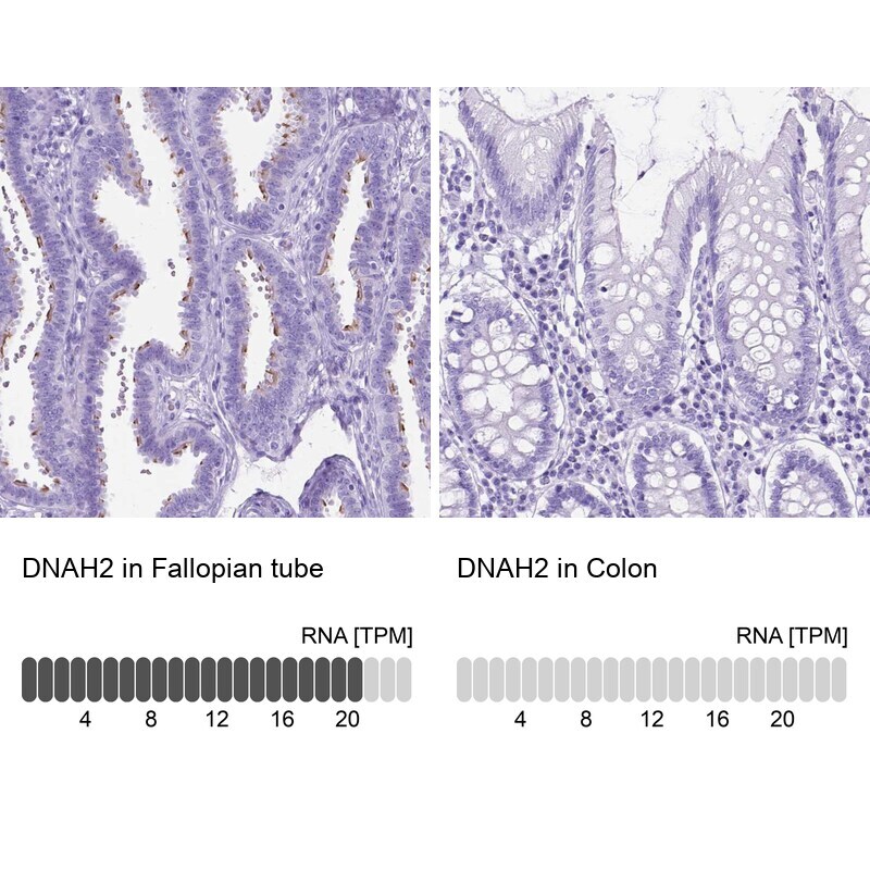
- Immunohistochemical analysis of DNAH2 in human fallopian tube using DNAH2 Polyclonal Antibody (Product # PA5-64309) shows high expression.
- Submitted by
- Invitrogen Antibodies (provider)
- Main image
- Experimental details
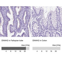
- Immunohistochemical staining of DNAH2 in human fallopian tube and colon tissues using DNAH2 Polyclonal Antibody (Product # PA5-64309). Corresponding DNAH2 RNA-seq data are presented for the same tissues.
- Submitted by
- Invitrogen Antibodies (provider)
- Main image
- Experimental details
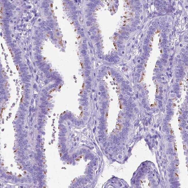
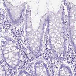
- Immunohistochemical staining of DNAH2 in human colon using DNAH2 Polyclonal Antibody (Product # PA5-64309) shows low expression as expected.

Supportive validation
- Submitted by
- Invitrogen Antibodies (provider)
- Main image
- Experimental details
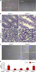
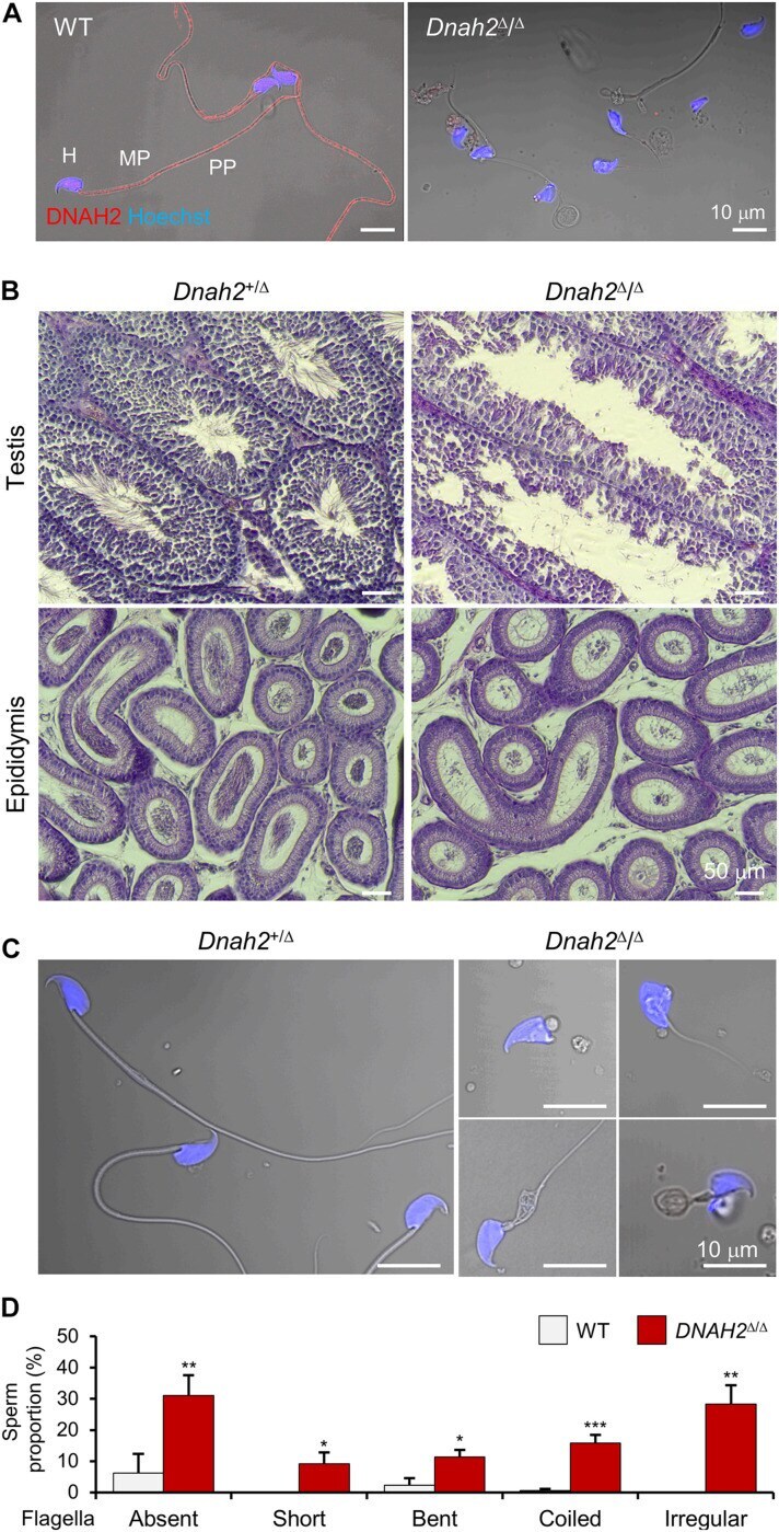
- FIGURE 2 DNAH2 deficiency in mice recapitulates multiple morphological abnormalities of the flagella in humans. (A) Confocal images of DNAH2 in WT and Dnah2 Delta/Delta sperm. DNAH2 were immunodetected from WT sperm but not from Dnah2 Delta/Delta sperm. Sperm heads were counterstained with Hoechst. Shown images are confocal images merged to the corresponding DIC images. H, head; MP, midpiece; PP, principal piece. (B) Testicular and epididymal histology of Dnah2 Delta/Delta mouse testis and epididymis. H/E stained sections show Dnah2 Delta/Delta males produce less sperm with elongated tails in the lumen of seminiferous tubule in testis ( top ), resulting in fewer sperm cells in the epididymis ( bottom , corpus). (C,D) Multiple morphological defects in epididymal Dnah2 Delta/Delta sperm. (C) Morphology of Dnah2 +/Delta ( left ) and Dnah2 Delta/Delta ( right ) sperm. Sperm from heterozygous males are morphologically normal. Dnah2 Delta/Delta sperm, however, present MMAF-like phenotypes, such as absent, short, coiled, and irregular-caliber flagella (from left top , clockwise direction). Sperm heads were Hoechst-stained (blue). Fluorescence confocal and corresponding DIC images are merged. (D) Proportions of sperm with defective flagellar morphologies. Sperm with absent, short, bent, coiled, and irregular-caliber flagella were quantified from WT (gray) and Dnah2 Delta/Delta (red) male mice ( N = 3, each). Sperm proportions of each pattern were statistically compared between WT and
- Submitted by
- Invitrogen Antibodies (provider)
- Main image

- Experimental details
- FIGURE 3 Flagellar ultrastructure is disorganized, and axonemal proteins express abnormally in Dnah2 - sperm. (A,B) Scanning electron microscopy images of Dnah2 +/Delta (A) and Dnah2 Delta/Delta (B) sperm. Dnah2 Delta/Delta sperm display absence of ( left ) or an irregular ( right ) mitochondrial helical sheath in the midpiece of the sperm tail, along with abnormal head shapes. Arrowheads indicate annulus. H, head; MP, midpiece; and PP, principal piece. (C,D) Transmission electron microscopy images of Dnah2 +/Delta (C) and Dnah2 Delta/Delta (D) sperm. Representative cross-section images of TEM reveal conformational defects in the midpiece of Dnah2 Delta/Delta sperm. Incomplete 9+2 axonemal structure ( left ), delocalized outer dense fibers (ODF, arrow) and microtubule doublet ( middle , arrowhead), and lack of the axonemal components ( right ) were observed in the Dnah2 Delta/Delta sperm. Inner (yellow asterisk) and outer (white asterisk) dynein arm structure were observed from microtubule doublet occasionally ( left , inset; scale bar = 50 nm). Microtubule doublets are numbered with the absence of the corresponding microtubule doublet in red. M, mitochondria. (E) Confocal fluorescence images of axonemal proteins in WT and Dnah2 Delta/Delta sperm. Dnah2 Delta/Delta sperm do not express another IDA component (DNAH1, magenta) and a radial spoke protein (RSPH3, green) but an ODA protein (DNAH9, yellow). Flagella microtubules (AcTub, white) and sperm heads (Hoechst, blue) ser
