 Explore
 Validate
 Learn
Learn Western blot
 Immunocytochemistry
Antibody data
- Antibody Data
- Antigen structure
- References [9]
- Comments [0]
- Validations
- Western blot [2]
- Immunohistochemistry [1]
Submit
Validation data
Reference
Comment
Report error
- Product number
- AF3264 - Provider product page

- Provider
- Novus Biologicals
- Product name
- Goat Polyclonal Cadherin-13 Antibody
- Antibody type
- Polyclonal
- Description
- Immunogen affinity purified. Detects human Cadherin-13 in direct ELISAs and Western blots. In direct ELISAs, approximately 100% cross-reactivity with recombinant mouse Cadherin-13 is observed, and less than 10% cross-reactivity with recombinant human (rh) N-Cadherin is observed, and less than 2% cross-reactivity with rhCadherin-8, rhCadherin-11, rhCadherin-17, rhE-Cadherin, rhP-Cadherin, rhVE-Cadherin and rhR-Cadherin is observed.
- Reactivity
- Human
- Host
- Goat
- Conjugate
- Unconjugated
- Isotype
- IgG
- Vial size
- 100 ug
- Concentration
- LYOPH
- Storage
- Use a manual defrost freezer and avoid repeated freeze-thaw cycles. 12 months from date of receipt, -20 to -70 degreesC as supplied. 1 month, 2 to 8 degreesC under sterile conditions after reconstitution. 6 months, -20 to -70 degreesC under sterile conditions after reconstitution.
Submitted references Pioglitazone strengthen therapeutic effect of adipose-derived regenerative cells against ischemic cardiomyopathy through enhanced expression of adiponectin and modulation of macrophage phenotype.
Actin cytoskeleton regulates functional anchorage-migration switch during T-cadherin-induced phenotype modulation of vascular smooth muscle cells.
Adiponectin/T-cadherin system enhances exosome biogenesis and decreases cellular ceramides by exosomal release.
The unique prodomain of T-cadherin plays a key role in adiponectin binding with the essential extracellular cadherin repeats 1 and 2.
Vagal nerve stimulation modifies neuronal activity and the proteome of excitatory synapses of amygdala/piriform cortex.
T-Cadherin Expression in the Epidermis and Adnexal Structures of Normal Skin.
Cross-talk between EGFR and T-cadherin: EGFR activation promotes T-cadherin localization to intercellular contacts.
T-cadherin is essential for adiponectin-mediated revascularization.
Identification of proteins associating with glycosylphosphatidylinositol- anchored T-cadherin on the surface of vascular endothelial cells: role for Grp78/BiP in T-cadherin-dependent cell survival.
Mori D, Miyagawa S, Matsuura R, Sougawa N, Fukushima S, Ueno T, Toda K, Kuratani T, Tomita K, Maeda N, Shimomura I, Sawa Y
Cardiovascular diabetology 2019 Mar 22;18(1):39
Cardiovascular diabetology 2019 Mar 22;18(1):39
Actin cytoskeleton regulates functional anchorage-migration switch during T-cadherin-induced phenotype modulation of vascular smooth muscle cells.
Frismantiene A, Kyriakakis E, Dasen B, Erne P, Resink TJ, Philippova M
Cell adhesion & migration 2018 Jan 2;12(1):69-85
Cell adhesion & migration 2018 Jan 2;12(1):69-85
Adiponectin/T-cadherin system enhances exosome biogenesis and decreases cellular ceramides by exosomal release.
Obata Y, Kita S, Koyama Y, Fukuda S, Takeda H, Takahashi M, Fujishima Y, Nagao H, Masuda S, Tanaka Y, Nakamura Y, Nishizawa H, Funahashi T, Ranscht B, Izumi Y, Bamba T, Fukusaki E, Hanayama R, Shimada S, Maeda N, Shimomura I
JCI insight 2018 Apr 19;3(8)
JCI insight 2018 Apr 19;3(8)
The unique prodomain of T-cadherin plays a key role in adiponectin binding with the essential extracellular cadherin repeats 1 and 2.
Fukuda S, Kita S, Obata Y, Fujishima Y, Nagao H, Masuda S, Tanaka Y, Nishizawa H, Funahashi T, Takagi J, Maeda N, Shimomura I
The Journal of biological chemistry 2017 May 12;292(19):7840-7849
The Journal of biological chemistry 2017 May 12;292(19):7840-7849
Vagal nerve stimulation modifies neuronal activity and the proteome of excitatory synapses of amygdala/piriform cortex.
Alexander GM, Huang YZ, Soderblom EJ, He XP, Moseley MA, McNamara JO
Journal of neurochemistry 2017 Feb;140(4):629-644
Journal of neurochemistry 2017 Feb;140(4):629-644
T-Cadherin Expression in the Epidermis and Adnexal Structures of Normal Skin.
Buechner S, Erne P, Resink TJ
Dermatopathology (Basel, Switzerland) 2016 Sep-Dec;3(4):68-78
Dermatopathology (Basel, Switzerland) 2016 Sep-Dec;3(4):68-78
Cross-talk between EGFR and T-cadherin: EGFR activation promotes T-cadherin localization to intercellular contacts.
Kyriakakis E, Maslova K, Frachet A, Ferri N, Contini A, Pfaff D, Erne P, Resink TJ, Philippova M
Cellular signalling 2013 May;25(5):1044-53
Cellular signalling 2013 May;25(5):1044-53
T-cadherin is essential for adiponectin-mediated revascularization.
Parker-Duffen JL, Nakamura K, Silver M, Kikuchi R, Tigges U, Yoshida S, Denzel MS, Ranscht B, Walsh K
The Journal of biological chemistry 2013 Aug 23;288(34):24886-97
The Journal of biological chemistry 2013 Aug 23;288(34):24886-97
Identification of proteins associating with glycosylphosphatidylinositol- anchored T-cadherin on the surface of vascular endothelial cells: role for Grp78/BiP in T-cadherin-dependent cell survival.
Philippova M, Ivanov D, Joshi MB, Kyriakakis E, Rupp K, Afonyushkin T, Bochkov V, Erne P, Resink TJ
Molecular and cellular biology 2008 Jun;28(12):4004-17
Molecular and cellular biology 2008 Jun;28(12):4004-17
No comments: Submit comment
Supportive validation
- Submitted by
- Novus Biologicals (provider)
- Main image
- Experimental details
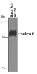
- Detection of Human Cadherin-13 by Western Blot. Western blot shows lysates of human brain (cortex) tissue. PVDF Membrane was probed with 1 µg/mL of Goat Anti-Human Cadherin-13 Antigen Affinity-purified Polyclonal Antibody (Catalog # AF3264) followed by HRP-conjugated Anti-Goat IgG Secondary Antibody (Catalog # HAF019). A specific band was detected for Cadherin-13 at approximately 105 kDa (as indicated). This experiment was conducted under reducing conditions and using Immunoblot Buffer Group 8.

- Submitted by
- Novus Biologicals (provider)
- Main image

- Experimental details
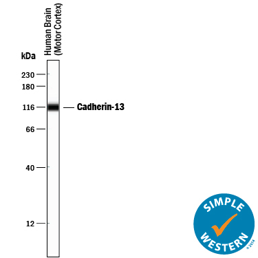
- Detection of Human Cadherin-13 by Simple WesternTM. Simple Western lane view shows lysates of human brain (motor cortex) tissue, loaded at 0.2 mg/mL. A specific band was detected for Cadherin-13 at approximately 114 kDa (as indicated) using 10 µg/mL of Goat Anti-Human Cadherin-13 Antigen Affinity-purified Polyclonal Antibody (Catalog # AF3264) followed by 1:50 dilution of HRP-conjugated Anti-Goat IgG Secondary Antibody (Catalog # HAF109). This experiment was conducted under reducing conditions and using the 12-230 kDa separation system.
Supportive validation
- Submitted by
- Novus Biologicals (provider)
- Main image
- Experimental details
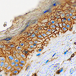
- Cadherin-13 in Human Skin. Cadherin-13 was detected in immersion fixed paraffin-embedded sections of human skin using Goat Anti-Human Cadherin-13 Antigen Affinity-purified Polyclonal Antibody (Catalog # AF3264) at 0.1 µg/mL overnight at 4 °C. Tissue was stained using the Anti-Goat HRP-DAB Cell & Tissue Staining Kit (brown; Catalog # CTS008) and counterstained with hematoxylin (blue). Specific staining was localized to plasma membranes of keratinocytes. View our protocol for Chromogenic IHC Staining of Paraffin-embedded Tissue Sections.
