 Explore
 Validate
 Learn
Learn Western blot
 Immunohistochemistry
Antibody data
- Antibody Data
- Antigen structure
- References [7]
- Comments [0]
- Validations
- Immunohistochemistry [1]
Submit
Validation data
Reference
Comment
Report error
- Product number
- HPA023719 - Provider product page

- Provider
- Atlas Antibodies
- Proper citation
- Atlas Antibodies Cat#HPA023719, RRID:AB_1856124
- Product name
- Anti-ESRP1
- Antibody type
- Polyclonal
- Description
- Polyclonal Antibody against Human ESRP1, Gene description: epithelial splicing regulatory protein 1, Alternative Gene Names: FLJ20171, RBM35A, Validated applications: IHC, WB, Uniprot ID: Q6NXG1, Storage: Store at +4°C for short term storage. Long time storage is recommended at -20°C.
- Reactivity
- Human
- Host
- Rabbit
- Conjugate
- Unconjugated
- Isotype
- IgG
- Vial size
- 100 µl
- Concentration
- 0.1 mg/ml
- Storage
- Store at +4°C for short term storage. Long time storage is recommended at -20°C.
- Handling
- The antibody solution should be gently mixed before use.
Submitted references Presence of spontaneous epithelial-mesenchymal plasticity in esophageal cancer
Role of PKCε in the epithelial-mesenchymal transition induced by FGFR2 isoform switch
Cell Plasticity-Related Phenotypes and Taxanes Resistance in Castration-Resistant Prostate Cancer
The Aberrant Expression of the Mesenchymal Variant of FGFR2 in the Epithelial Context Inhibits Autophagy
Androgen-regulated transcription of ESRP2 drives alternative splicing patterns in prostate cancer
Esrp1 is a marker of mouse fetal germ cells and differentially expressed during spermatogenesis
Epithelial Splicing Regulatory Proteins 1 (ESRP1) and 2 (ESRP2) Suppress Cancer Cell Motility via Different Mechanisms
Tsuchihashi K, Hirata Y, Yamasaki J, Suina K, Tanoue K, Yae T, Masuda K, Baba E, Akashi K, Kitagawa Y, Saya H, Nagano O
Biochemistry and Biophysics Reports 2022;30
Biochemistry and Biophysics Reports 2022;30
Role of PKCε in the epithelial-mesenchymal transition induced by FGFR2 isoform switch
Ranieri D, Nanni M, Persechino F, Torrisi M, Belleudi F
Cell Communication and Signaling 2020;18(1)
Cell Communication and Signaling 2020;18(1)
Cell Plasticity-Related Phenotypes and Taxanes Resistance in Castration-Resistant Prostate Cancer
Jiménez N, Reig Ò, Montalbo R, Milà-Guasch M, Nadal-Dieste L, Castellano G, Lozano J, Victoria I, Font A, Rodriguez-Vida A, Carles J, Suárez C, Domènech M, Sala-González N, Fernández P, Rodríguez-Carunchio L, Díaz S, Prat A, Marín-Aguilera M, Mellado B
Frontiers in Oncology 2020;10
Frontiers in Oncology 2020;10
The Aberrant Expression of the Mesenchymal Variant of FGFR2 in the Epithelial Context Inhibits Autophagy
Nanni M, Ranieri D, Persechino F, Torrisi M, Belleudi F
Cells 2019;8(7):653
Cells 2019;8(7):653
Androgen-regulated transcription of ESRP2 drives alternative splicing patterns in prostate cancer
Munkley J, Li L, Krishnan S, Hysenaj G, Scott E, Dalgliesh C, Oo H, Maia T, Cheung K, Ehrmann I, Livermore K, Zielinska H, Thompson O, Knight B, McCullagh P, McGrath J, Crundwell M, Harries L, Daugaard M, Cockell S, Barbosa-Morais N, Oltean S, Elliott D
eLife 2019;8
eLife 2019;8
Esrp1 is a marker of mouse fetal germ cells and differentially expressed during spermatogenesis
Singh S, Saeidi S, Shapouri F, de Iongh R, Casagranda F, Sutherland J, Western P, McLaughlin E, Familari M, Hime G
PLOS ONE 2018;13(1):e0190925
PLOS ONE 2018;13(1):e0190925
Epithelial Splicing Regulatory Proteins 1 (ESRP1) and 2 (ESRP2) Suppress Cancer Cell Motility via Different Mechanisms
Ishii H, Saitoh M, Sakamoto K, Kondo T, Katoh R, Tanaka S, Motizuki M, Masuyama K, Miyazawa K
Journal of Biological Chemistry 2014;289(40):27386-27399
Journal of Biological Chemistry 2014;289(40):27386-27399
No comments: Submit comment
Supportive validation
- Submitted by
- Atlas Antibodies (provider)
- Enhanced method
- Orthogonal validation
- Main image
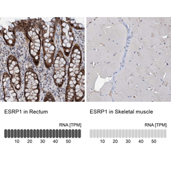
- Experimental details
- Immunohistochemistry analysis in human rectum and skeletal muscle tissues using HPA023719 antibody. Corresponding ESRP1 RNA-seq data are presented for the same tissues.
- Sample type
- Human
- Protocol
- Protocol
