 Explore
 Validate
 Learn
Learn Immunocytochemistry
 Immunoprecipitation
Antibody data
- Antibody Data
- Antigen structure
- References [4]
- Comments [0]
- Validations
- Immunocytochemistry [1]
- Flow cytometry [3]
Submit
Validation data
Reference
Comment
Report error
- Product number
- MA1-81930 - Provider product page

- Provider
- Invitrogen Antibodies
- Product name
- CD90 Monoclonal Antibody (F15-42-1)
- Antibody type
- Monoclonal
- Antigen
- Purifed from natural sources
- Description
- The epitope recognized by this antibody is reported to be sensitive to formaldehyde fixation and tissue processing. We recommend the use of acetone fixation for frozen sections. A suggested positive control for immunohistochemical applications is human brain or thymus. For FACS analysis, use 10 µL of the suggested working dilution to label 1x10^6 cells in 100 µL. Mouse anti Human CD90 antibody, clone F15-42-1 recognizes the human CD90 cell surface antigen, a approximately 25 kDa glycoprotein homologous to rat Thy1. Mouse anti Human CD90 antibody, clone F15-42-1 recognizes the human CD90 cell surface antigen, a approximately 25 kDa glycoprotein homologous to rat Thy1.
- Reactivity
- Human
- Host
- Mouse
- Isotype
- IgG
- Antibody clone number
- F15-42-1
- Vial size
- 20 µg
- Concentration
- 1 mg/mL
- Storage
- Store at 4°C short term. For long term storage, store at -20°C, avoiding freeze/thaw cycles.
Submitted references Quantification of Antibody Persistence for Cell Surface Protein Labeling.
GLI1 and AXIN2 Are Distinctive Markers of Human Calvarial Mesenchymal Stromal Cells in Nonsyndromic Craniosynostosis.
Corneal keratocyte transition to mesenchymal stem cell phenotype and reversal using serum-free medium supplemented with fibroblast growth factor-2, transforming growth factor-β3 and retinoic acid.
Effect of culture medium on propagation and phenotype of corneal stroma-derived stem cells.
Dempsey ME, Woodford-Berry O, Darling EM
Cellular and molecular bioengineering 2021 Jun;14(3):267-277
Cellular and molecular bioengineering 2021 Jun;14(3):267-277
GLI1 and AXIN2 Are Distinctive Markers of Human Calvarial Mesenchymal Stromal Cells in Nonsyndromic Craniosynostosis.
Di Pietro L, Barba M, Prampolini C, Ceccariglia S, Frassanito P, Vita A, Guadagni E, Bonvissuto D, Massimi L, Tamburrini G, Parolini O, Lattanzi W
International journal of molecular sciences 2020 Jun 19;21(12)
International journal of molecular sciences 2020 Jun 19;21(12)
Corneal keratocyte transition to mesenchymal stem cell phenotype and reversal using serum-free medium supplemented with fibroblast growth factor-2, transforming growth factor-β3 and retinoic acid.
Sidney LE, Hopkinson A
Journal of tissue engineering and regenerative medicine 2018 Jan;12(1):e203-e215
Journal of tissue engineering and regenerative medicine 2018 Jan;12(1):e203-e215
Effect of culture medium on propagation and phenotype of corneal stroma-derived stem cells.
Sidney LE, Branch MJ, Dua HS, Hopkinson A
Cytotherapy 2015 Dec;17(12):1706-22
Cytotherapy 2015 Dec;17(12):1706-22
No comments: Submit comment
Supportive validation
- Submitted by
- Invitrogen Antibodies (provider)
- Main image

- Experimental details
- Immunofluorescence analysis of CD90 was performed using 70% confluent log phase SH-SY5Y cells. The cells were fixed with 4% paraformaldehyde for 10 minutes, permeabilized with 0.1% Triton™ X-100 for 15 minutes, and blocked with 2% BSA for 45 minutes at room temperature. The cells were labeled with CD90 Monoclonal Antibody (F15-42-1) (Product # MA5-16671) at 1:100 dilution in 0.1% BSA, incubated at 4 degree celsius overnight and then labeled with Donkey anti-Rabbit IgG (H+L) Highly Cross-Adsorbed Secondary Antibody, Alexa Fluor Plus 488 (Product # A32790), (1:2000 dilution), for 45 minutes at room temperature (Panel a: Green). Nuclei (Panel b: Green) were stained with ProLong™ Diamond Antifade Mountant with DAPI (Product # P36962). F-actin (Panel c: Red) was stained with Rhodamine Phalloidin (Product # R415, 1:300 dilution). Panel d represents the merged image showing membranous and cytosolic localization. Panel e represents MCF7 cells showing low expression of CD90. Panel f represents control cells with no primary antibody to assess background. The images were captured at 60X magnification.

Supportive validation
- Submitted by
- Invitrogen Antibodies (provider)
- Main image
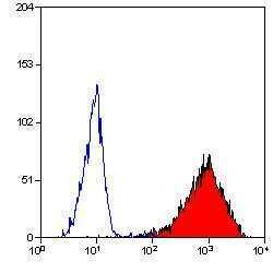
- Experimental details
- Flow cytometric analysis of HUT78 T monocytes using a CD90/Thy-1 monoclonal antibody (Product # MA5-16671)

- Submitted by
- Invitrogen Antibodies (provider)
- Main image
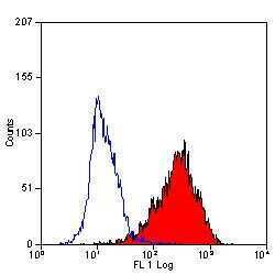
- Experimental details
- Flow cytometric analysis of HUT78 cells using a CD90/Thy-1 monoclonal antibody (Product # MA5-16671)

- Submitted by
- Invitrogen Antibodies (provider)
- Main image
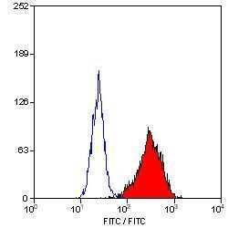
- Experimental details
- Flow cytometric analysis of HUT78 T cells using a CD90/Thy-1 monoclonal antibody (Product # MA5-16671)
