Explore
 Validate
 Learn
Learn Flow cytometry
Antibody data
- Antibody Data
- Antigen structure
- References [3]
- Comments [0]
- Validations
- Flow cytometry [1]
Submit
Validation data
Reference
Comment
Report error
- Product number
- MAB5958 - Provider product page

- Provider
- Abnova Corporation
- Proper citation
- Abnova Corporation Cat#MAB5958, RRID:AB_10554784
- Product name
- Thy1 monoclonal antibody, clone 30-H12 (APC)
- Antibody type
- Monoclonal
- Description
- Rat monoclonal antibody raised against native Thy1.
- Isotype
- IgG
- Antibody clone number
- 30-H12
- Storage
- Store in the dark at 4°C. Do not freeze.Avoid prolonged exposure to light.Aliquot to avoid repeated freezing and thawing.
Submitted references Complete structure of the glycosyl phosphatidylinositol membrane anchor of rat brain Thy-1 glycoprotein.
Neuronal cell Thy-1 glycoprotein: homology with immunoglobulin.
T cell subsets defined by expression of Lyt-1,2,3 and Thy-1 antigens. Two-parameter immunofluorescence and cytotoxicity analysis with monoclonal antibodies modifies current views.
Homans SW, Ferguson MA, Dwek RA, Rademacher TW, Anand R, Williams AF
Nature 1988 May 19;333(6170):269-72
Nature 1988 May 19;333(6170):269-72
Neuronal cell Thy-1 glycoprotein: homology with immunoglobulin.
Williams AF, Gagnon J
Science (New York, N.Y.) 1982 May 14;216(4547):696-703
Science (New York, N.Y.) 1982 May 14;216(4547):696-703
T cell subsets defined by expression of Lyt-1,2,3 and Thy-1 antigens. Two-parameter immunofluorescence and cytotoxicity analysis with monoclonal antibodies modifies current views.
Ledbetter JA, Rouse RV, Micklem HS, Herzenberg LA
The Journal of experimental medicine 1980 Aug 1;152(2):280-95
The Journal of experimental medicine 1980 Aug 1;152(2):280-95
No comments: Submit comment
Supportive validation
- Submitted by
- Abnova Corporation (provider)
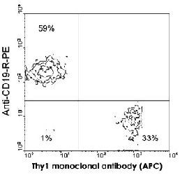
- Main image

- Experimental details
- BALB/c splenocytes were double-stained with Thy1 monoclonal antibody, clone 30-H12 (APC) (Cat # MAB5958) and Cd19 monoclonal antibody, clone 6D5 (PE) (Cat # MAB5689). Small lymphocytes were then gated and analyzed on a FACScan™ flow cytometer (BDIS, San Jose, CA).
- Validation comment
- Flow Cytometry