 Explore
 Validate
 Learn
Learn Western blot
Antibody data
- Antibody Data
- Antigen structure
- References [2]
- Comments [0]
- Validations
- Western blot [1]
- Immunocytochemistry [3]
- Immunohistochemistry [1]
- Flow cytometry [3]
Submit
Validation data
Reference
Comment
Report error
- Product number
- AF2067 - Provider product page

- Provider
- R&D Systems
- Product name
- Human/Porcine/Canine CD90/Thy1 Antibody
- Antibody type
- Polyclonal
- Description
- Immunogen affinity purified. Detects human CD90/Thy1 in direct ELISAs and Western blots. In direct ELISAs, approximately 50% cross-reactivity with recombinant mouse CD90 is observed.
- Reactivity
- Human, Canine, Porcine
- Host
- Sheep
- Conjugate
- Unconjugated
- Antigen sequence
P04216- Isotype
- IgG
- Vial size
- 100 ug
- Concentration
- LYOPH
- Storage
- Use a manual defrost freezer and avoid repeated freeze-thaw cycles. 12 months from date of receipt, -20 to -70 °C as supplied. 1 month, 2 to 8 °C under sterile conditions after reconstitution. 6 months, -20 to -70 °C under sterile conditions after reconstitution.
Submitted references Podoplanin regulates the migration of mesenchymal stromal cells and their interaction with platelets.
Pathological Remodeling of Mitral Valve Leaflets from Unphysiologic Leaflet Mechanics after Undersized Mitral Annuloplasty to Repair Ischemic Mitral Regurgitation.
Ward LSC, Sheriff L, Marshall JL, Manning JE, Brill A, Nash GB, McGettrick HM
Journal of cell science 2019 Feb 25;132(5)
Journal of cell science 2019 Feb 25;132(5)
Pathological Remodeling of Mitral Valve Leaflets from Unphysiologic Leaflet Mechanics after Undersized Mitral Annuloplasty to Repair Ischemic Mitral Regurgitation.
Sielicka A, Sarin EL, Shi W, Sulejmani F, Corporan D, Kalra K, Thourani VH, Sun W, Guyton RA, Padala M
Journal of the American Heart Association 2018 Nov 6;7(21):e009777
Journal of the American Heart Association 2018 Nov 6;7(21):e009777
No comments: Submit comment
Supportive validation
- Submitted by
- R&D Systems (provider)
- Main image
- Experimental details
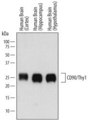
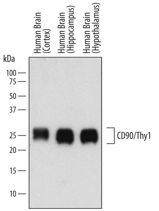
- Detection of Human CD90/Thy1 by Western Blot. Western blot shows lysates of human brain (cortex) tissue, human brain (hippocampus) tissue, and human brain (hypothalamus) tissue. PVDF membrane was probed with 1 µg/mL of Sheep Anti-Human/Porcine/Canine CD90/Thy1 Antigen Affinity-purified Polyclonal Antibody (Catalog # AF2067) followed by HRP-conjugated Anti-Sheep IgG Secondary Antibody (Catalog # HAF016). A specific band was detected for CD90/Thy1 at approximately 22-30 kDa (as indicated). This experiment was conducted under reducing conditions and using Immunoblot Buffer Group 1.
Supportive validation
- Submitted by
- R&D Systems (provider)
- Main image
- Experimental details
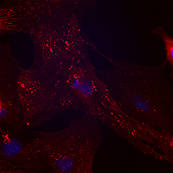
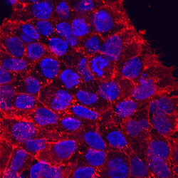
- CD90/Thy1 in Porcine Mesenchymal Stem Cells. CD90/Thy1 was detected in immersion fixed porcine mesenchymal stem cells using Sheep Anti-Human/Porcine/Canine CD90/Thy1 Antigen Affinity-purified Polyclonal Antibody (Catalog # AF2067) at 10 µg/mL for 3 hours at room temperature. Cells were stained using the NorthernLights™ 557-conjugated Anti-Sheep IgG Secondary Antibody (red; Catalog # NL010) and counterstained with DAPI (blue). Specific staining was localized to cell surfaces and cytoplasm. View our protocol for Fluorescent ICC Staining of Stem Cells on Coverslips.
- Submitted by
- R&D Systems (provider)
- Main image
- Experimental details
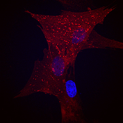
- CD90/Thy1 in BG01V Human Embyonic Stem Cells. CD90/Thy1 was detected in immersion fixed BG01V human embryonic stem cells using Sheep Anti-Human/Porcine/Canine CD90/Thy1 Antigen Affinity-purified Polyclonal Antibody (Catalog # AF2067) at 10 µg/mL for 3 hours at room temperature. Cells were stained using the NorthernLights™ 557-conjugated Anti-Sheep IgG Secondary Antibody (red; Catalog # NL010) and counterstained with DAPI (blue). Specific staining was localized to cytoplasm. View our protocol for Fluorescent ICC Staining of Stem Cells on Coverslips.

- Submitted by
- R&D Systems (provider)
- Main image
- Experimental details
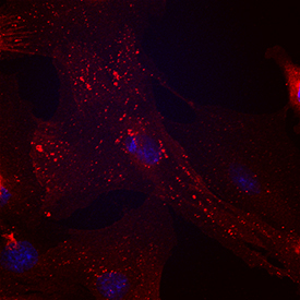
- CD90/Thy1 in Canine Mesenchymal Stem Cells. CD90/Thy1 was detected in immersion fixed canine mesenchymal stem cells using Sheep Anti-Human/Porcine/Canine CD90/Thy1 Antigen Affinity-purified Polyclonal Antibody (Catalog # AF2067) at 10 µg/mL for 3 hours at room temperature. Cells were stained using the NorthernLights™ 557-conjugated Anti-Sheep IgG Secondary Antibody (red; Catalog # NL010) and counterstained with DAPI (blue). Specific staining was localized to cell surfaces. View our protocol for Fluorescent ICC Staining of Stem Cells on Coverslips.

Supportive validation
- Submitted by
- R&D Systems (provider)
- Main image
- Experimental details
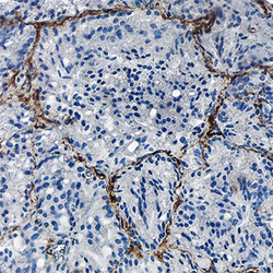
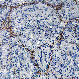
- CD90/Thy1 in Human Prostate Cancer Tissue. CD90/Thy1 was detected in formalin fixed paraffin-embedded sections of human prostate cancer tissue using Sheep Anti-Human/Porcine/Canine CD90/Thy1 Antigen Affinity-purified Polyclonal Antibody (Catalog # AF2067) at 1.7 µg/mL overnight at 4 °C. Tissue was stained using the Anti-Sheep HRP-DAB Cell & Tissue Staining Kit (brown; Catalog # CTS019) and counterstained with hematoxylin (blue). Specific staining was localized to endothelial cells. View our protocol for Chromogenic IHC Staining of Paraffin-embedded Tissue Sections.
Supportive validation
- Submitted by
- R&D Systems (provider)
- Main image

- Experimental details
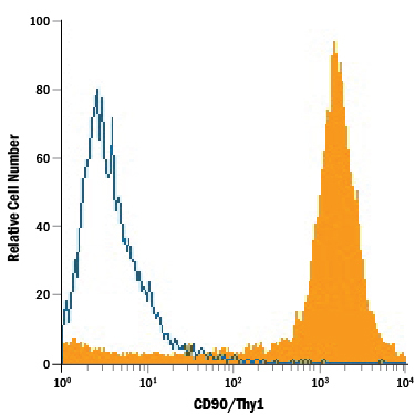
- Detection of CD90/Thy1 in Porcine Mesenchymal Stem Cells by Flow Cytometry. Porcine mesenchymal stem cells were stained with Sheep Anti-Human/Porcine/Canine CD90/Thy1 Antigen Affinity-purified Polyclonal Antibody (Catalog # AF2067, filled histogram) or isotype control antibody (Catalog # 5-001-A, open histogram), followed by PE-conjugated Anti-Sheep IgG Secondary Antibody (Catalog # F0126).
- Submitted by
- R&D Systems (provider)
- Main image

- Experimental details
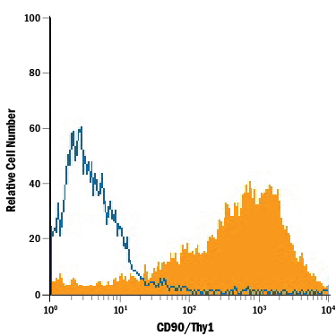
- Detection of CD90/Thy1 in Canine Mesenchymal Stem Cells by Flow Cytometry. Canine mesenchymal stem cells were stained with Sheep Anti-Human/Porcine/Canine CD90/Thy1 Antigen Affinity-purified Polyclonal Antibody (Catalog # AF2067, filled histogram) or isotype control antibody (Catalog # 5-001-A, open histogram), followed by PE-conjugated Anti-Sheep IgG Secondary Antibody (Catalog # F0126).
- Submitted by
- R&D Systems (provider)
- Main image
- Experimental details
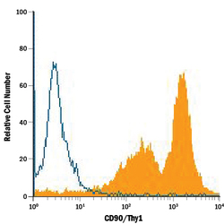
- Detection of CD90/Thy1 in Human Mesenchymal Stem Cells by Flow Cytometry. Human mesenchymal stem cells were stained with Sheep Anti-Human/Porcine/Canine CD90/Thy1 Antigen Affinity-purified Polyclonal Antibody (Catalog # AF2067, filled histogram) or isotype control antibody (Catalog # 5-001-A, open histogram), followed by PE-conjugated Anti-Sheep IgG Secondary Antibody (Catalog # F0126).
