Explore
 Validate
 Learn
Learn Immunocytochemistry
 Immunohistochemistry
Antibody data
- Antibody Data
- Antigen structure
- References [3]
- Comments [0]
- Validations
- Immunocytochemistry [1]
Submit
Validation data
Reference
Comment
Report error
- Product number
- AMAb91361 - Provider product page

- Provider
- Atlas Antibodies
- Proper citation
- Atlas Antibodies Cat#AMAb91361, RRID:AB_2716651
- Product name
- Anti-FOXP2
- Antibody type
- Monoclonal
- Description
- Monoclonal Antibody against Human FOXP2, Clone ID: CL5310, Gene description: forkhead box P2, Alternative Gene Names: CAGH44, SPCH1, TNRC10, Validated applications: IHC, ICC, Uniprot ID: O15409, Storage: Store at +4°C for short term storage. Long time storage is recommended at -20°C.
- Reactivity
- Human, Mouse
- Host
- Mouse
- Conjugate
- Unconjugated
- Isotype
- IgG
- Antibody clone number
- CL5310
- Vial size
- 100 µl
- Concentration
- 0.7 mg/ml
- Storage
- Store at +4°C for short term storage. Long time storage is recommended at -20°C.
- Handling
- The antibody solution should be gently mixed before use.
Submitted references Dissection of insular cortex layer 5 reveals two sublayers with opposing modulatory roles in appetitive drinking behavior.
A Ctnnb1 enhancer regulates neocortical neurogenesis by controlling the abundance of intermediate progenitors
Abnormal neocortex arealization and Sotos-like syndrome–associated behavior in Setd2 mutant mice
Takemoto M, Kato S, Kobayashi K, Song WJ
iScience 2023 Jun 16;26(6):106985
iScience 2023 Jun 16;26(6):106985
A Ctnnb1 enhancer regulates neocortical neurogenesis by controlling the abundance of intermediate progenitors
Wang J, Wang A, Tian K, Hua X, Zhang B, Zheng Y, Kong X, Li W, Xu L, Wang J, Li Z, Liu Y, Zhou Y
Cell Discovery 2022;8(1)
Cell Discovery 2022;8(1)
Abnormal neocortex arealization and Sotos-like syndrome–associated behavior in Setd2 mutant mice
Xu L, Zheng Y, Li X, Wang A, Huo D, Li Q, Wang S, Luo Z, Liu Y, Xu F, Wu X, Wu M, Zhou Y
Science Advances 2021;7(1)
Science Advances 2021;7(1)
No comments: Submit comment
Supportive validation
- Submitted by
- Atlas Antibodies (provider)
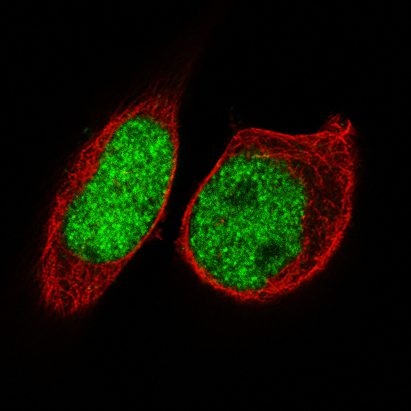
- Main image
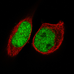
- Experimental details
- Immunofluorescence staining of RH-30 cells using the Anti-FOXP2 monoclonal antibody, showing specific staining in the nucleoplasm in green. Microtubule- and nuclear probes are visualized in red and blue, respectively (where available).
- Sample type
- Human